Upper third musculature — One elevator against four depressors
Frontalis, corrugator, procerus, depressor supercilii and orbicularis: origin, insertion, layer and innervation of each, the wrinkle each produces, and the exact translation into point, depth and dose.
Content intended for healthcare professionals. It does not replace clinical assessment and is not written as patient information.
Key points
- The whole upper third is one elevator against four depressors. Frontalis raises; corrugator, procerus, depressor supercilii and orbicularis lower.
- Hence the highest-yield manoeuvre in all of toxin work: blocking the depressors raises the brow without touching frontalis, and without freezing the forehead.
- Frontalis has no bony origin: it arises from the galea. That is why its tone depends on the whole scalp and why the brow follows the forehead.
- The corrugator changes depth along its course: deep at the origin, superficial at the insertion. One muscle, two needle depths.
- The arteries here are terminal branches of the ophthalmic, and therefore of the internal carotid. That is the anatomical reason for the risk of blindness when injecting filler.
The five muscles, one by one
| Muscle | From where to where | What it does and which wrinkle |
|---|---|---|
| Frontalis | From the galea — no bony origin — to the brow dermis, interdigitating with procerus, corrugator, depressor supercilii and orbicularis. It ends laterally at the temporal crest | The only elevator. Vertical fibres: it produces the horizontal forehead lines |
| Corrugator supercilii | From the medial supraorbital rim, on bone, to the dermis of the middle third of the brow, passing through frontalis and orbicularis | Draws the brow medially and down. Horizontal and oblique fibres: it produces the vertical glabellar lines |
| Procerus | From the fascia over the nasal bone and upper lateral cartilage to the glabellar dermis | Lowers the medial brow. Vertical fibres: it produces the horizontal line at the nasal root |
| Depressor supercilii | From the medial orbital rim, next to the lacrimal sac, to the medial brow dermis. Very superficial | Selectively depresses the brow head. Often forgotten, and responsible for the dropped medial brow |
| Orbicularis oculi | Three portions: orbital, palpebral — preseptal and pretarsal — and lacrimal. It encircles the orbit | The orbital part is voluntary closure and lateral brow depressor: it produces crow’s feet. The palpebral part is involuntary blinking |
Layer and innervation of each
| Muscle | Layer | Innervation |
|---|---|---|
| Frontalis | Layer 3, between the two galeal leaves | Temporal branch, on the deep surface |
| Corrugator | Deep at origin, superficial at insertion: it crosses layers | Temporal branch |
| Procerus | Layer 3, superficial | Classically described via the temporal branch, though some descriptions attribute it to the buccal branch |
| Depressor supercilii | Very superficial, almost subdermal | Temporal branch |
| Orbicularis | Layer 1, the most superficial of all | Temporal branch above and zygomatic below |
The consequence of frontalis having no bone
It is the only facial elevator not anchored to the skeleton: it arises from a mobile aponeurosis, the galea, which in turn continues into occipitalis behind and the superficial temporal fascia at the sides. Three consequences follow, used daily. One: for frontalis to elevate effectively the scalp must be under tension, which is why widely releasing the scalp in a brow lift not only avoids closure tension but changes the fulcrum. Two: since it ends at the temporal crest, it does not reach the brow tail, which is left without an elevator. Three: since it inserts into dermis interdigitating with the depressors, there is no clean boundary between elevating and depressing within the brow itself, which is why toxin diffuses so readily from one to the other.
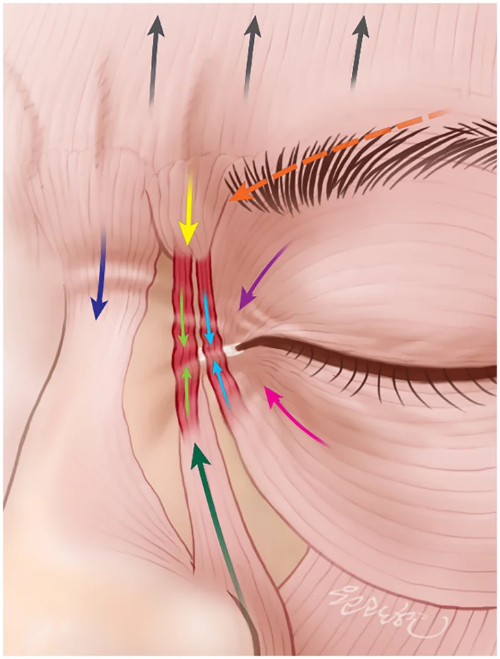
The glabellar complex: four muscles doing the same thing
Procerus, corrugators, depressor supercilii and the medial orbicularis work as a single depressor group over the brow head, which is why they are treated together. What differentiates them is the direction of their pull, and that direction is read in the wrinkle: procerus pulls straight down and leaves a horizontal line at the root; the corrugator pulls medially and leaves vertical lines. Understanding the group as one unit changes the consultation: glabellar treatment stopped being an independent indication and became part of harmonising brow shape and position.
The corrugator is two muscles as far as depth goes
It arises on bone and ends in dermis, so it rises through the planes along its course. At the medial origin it is deep and the needle must go in perpendicular and full depth; at the lateral insertion it is superficial and one third of the needle suffices. Injecting the lateral portion at the medial depth is precisely the manoeuvre that deposits product below the septum and produces eyelid ptosis. There is also an anatomical variant worth checking beforehand: there is a short corrugator and a long corrugator, and the latter reaches far more laterally than expected, so the vertical glabellar line may be produced by fibres lying outside the usual point.
Where to enter, how and how much
| Target | Technique and depth | Indicative dose |
|---|---|---|
| Frontalis | Pinch the skin until a papule appears and insert the needle angled upward, one third of its length | 1-2 U at 5 sites and 0.5-1 U at 2 optional sites. Mean dose 10 U, total under 20 U |
| Procerus | Pinch and insert half the needle, angled upward, centrally | Part of the five 4 U doses of the glabella, total 20 U |
| Medial corrugator | Pinch and insert the full needle, angled laterally and upward | 4 U per point, one point each side |
| Lateral corrugator | One third of the needle, angled laterally and upward: the muscle is more superficial here | 4 U per point, one point each side |
| Orbicularis, crow’s feet | One third of the needle or subdermal, very superficial. Eyes closed, needle always directed away from the eye and a finger protecting the eyelid | 4 U at 3 sites per side, total 24 U. In practice many drop to 2 U per point |
| Brow lifting | Glabellar points plus the lateral orbicularis: 3 to 4 sites per side | 2-3 U per site, 12 U per side. Overall total around 44 U |
| Eye aperture widening | A single point in orbicularis, very superficial | 1-2 U per side, total 2-4 U |
Rules that prevent the dropped brow
- Do not inject frontalis lateral to the mid-pupillary line. It is the rule that prevents most brow-tail ptosis.
- More dose above and less below on the forehead: the lower frontalis fibres are the ones holding the brow up.
- Extend the points far enough laterally so as not to leave a hyperactive lateral portion: that is what produces the peaked brow.
- To open the gaze: one point at the brow tail touching the orbital rim, and treat the corrugator without touching frontalis.
- If a comma-shaped line appears over the brow tail from a strong frontalis, 2 U very superficially there resolves it.
- Treating frontalis obliges also treating the glabellar depressors and periorbital lines: otherwise the brow drops.
Differences by sex and by forehead
The male brow is straight and over the rim; the female brow is arched, above the rim and peaking towards the lateral limbus. That changes the point pattern: in men the aim is maintaining horizontality, with points in parallel lines and around 4 U per point; in women a W pattern is used, respecting the arch. Total upper-third dose is around 50 U in men and 40 U in women. And one anatomical exception: in short foreheads it is wise to drop to 2 U per point, because the margin between relaxing and dropping the brow is far narrower.
One detail about crow’s feet
The area is richly vascularised and is where most bruising occurs, so it is worth seeing the superficial vessels before injecting and entering parallel and very superficially, at 1 or 2 mm. There is also a rarely cited precaution: in patients with eye bags or poor circulation in the tear trough, it is wise not to load the crow’s feet with points, because orbicularis contributes to lymphatic drainage of the area and blocking it makes this worse. And one warning always to give: treatment does not eliminate all the lines here, particularly those already static.
The vasculature, and why an eye can be lost here
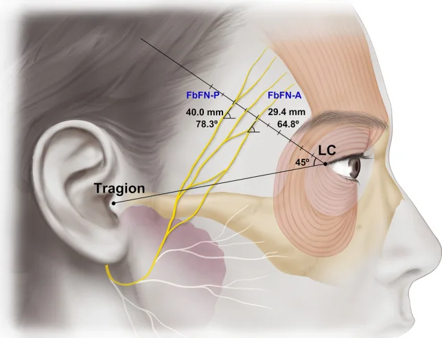
The key arteries of the upper third are the supraorbital and supratrochlear, which are terminal branches of the ophthalmic artery and therefore belong to the internal carotid territory; and the superficial temporal, a terminal branch of the external carotid. The consequence is direct: a filler embolus forced under pressure into the supratrochlear or supraorbital can travel retrograde to the central retinal artery and cause blindness. It is not a complication of technique: it is a consequence of anatomy, which is why precautions here are not optional. The nerves accompany the arteries: the supratrochlear runs with the corrugator and beneath the frontalis fascia to the medial and central forehead, and the supraorbital exits its foramen or notch and ascends beneath the same fascia to the anterolateral forehead and scalp.
Upper-face filler: where and in which plane
| Area | Technique | Volume and caution |
|---|---|---|
| Temple | Entry 1 cm above the lateral orbital rim and 1 cm lateral to the temporal crest. Needle perpendicular down to bone, aspirate, very slow supraperiosteal bolus | 0.5-1 ml per side, up to 2 ml in severe deficit. Avoid the subcutaneous superficial temporal artery and vein. Do not inject deeply in the lower or posterior fossa above the arch: internal maxillary branches are there, with a risk of palatal necrosis |
| Brow | Two supraperiosteal points, starting at the lateral end. Palpate the rim and protect with a finger so product does not migrate into the eyelid | 0.1 ml per site. Useful when toxin has not raised the tail enough. Avoid the supraorbital foramen and do not overcorrect: it gives a prominent brow and eyelid oedema |
| Forehead | Six points, three per side, always more than 2 cm from the brow. The tip must rest on bone, beneath the galea: that is the avascular plane. Aspirate before each point and massage at the end | Up to 0.1 ml per site. Avoid the supraorbital and supratrochlear bundles and the frontal branches of the superficial temporal. Massage is not optional: it is what gives a uniform contour |
Precautions that apply to any injection here
- BeforeRemove makeup, antisepsis with chlorhexidine, povidone or alcohol, and do not treat over irritated skin, active acne or infection
- NeedleChange it often: a blunt needle hurts more and contaminates more. Smaller gauges enter smaller vessels but force slower injection, which is what protects
- InjectingSlow, smooth and methodical, always. Aspirate before each bolus: it does not guarantee being outside a vessel, but it rules out many cases
- If in doubtIn areas of difficult anatomy, a blunt cannula instead of a needle: it reduces inadvertent intravascular injection
- If it blanchesVascular compromise: hyaluronidase in the area immediately, vigorous massage and warm compresses
- OrderIf in separate sessions, toxin first and filler after. If on the same day, filler first with its massage and then toxin
A low brow: whose problem is it?
Is the whole brow low or only the tail?
What this anatomy explains about the brow lift
The three decisions of brow surgery follow directly from this. Why the tail falls first: because frontalis ends at the temporal crest and only orbicularis remains there, and it depresses. Why release must precede traction: because corrugator and depressor supercilii anchor the medial brow to bone, and traction that does not free them merely tensions them. Why caution is needed in men: because a straight brow over the rim is a masculine feature and raising it feminises. Whoever holds the muscular map indicates better, and above all knows when the problem is solved with a syringe and when it is not.
How to document the session
- Video rather than a still: it is more dynamic and captures what a photograph misses. Gentle expression first, then forced.
- Three commands exploring the three groups: “smile showing your canines” for orbicularis, “raise your brows” for frontalis, “frown” for the glabella.
- Retouch at two weeks if needed, and do not repeat treatment before three months.
- Check and record before injecting: brow height, frontalis strength, skin elasticity and pre-existing asymmetries.
References
- 1.de Maio M, Swift A, Signorini M, Fagien S. Facial assessment and injection guide for botulinum toxin and injectable hyaluronic acid fillers: focus on the upper face. Plast Reconstr Surg. 2017;140(2):265e-276e.
- 2.Freilinger G, Gruber H, Happak W, Pechmann U. Surgical anatomy of the mimic muscle system and the facial nerve: importance for reconstructive and aesthetic surgery. Plast Reconstr Surg. 1987;80(5):686-690.
- 3.Knize DM. An anatomically based study of the mechanism of eyebrow ptosis. Plast Reconstr Surg. 1996;97(7):1321-1333.
- 4.Janis JE, Ghavami A, Lemmon JA, Leedy JE, Rohrich RJ. Anatomy of the corrugator supercilii muscle: part I. Corrugator topography. Plast Reconstr Surg. 2007;120(6):1647-1653.
- 5.Janis JE, Ghavami A, Lemmon JA, Leedy JE, Rohrich RJ. The anatomy of the corrugator supercilii muscle: part II. Supraorbital nerve branching patterns. Plast Reconstr Surg. 2008;121(1):233-240.
- 6.Knize DM. Transpalpebral approach to the corrugator supercilii and procerus muscles. Plast Reconstr Surg. 1995;95(1):52-60.
- 7.Khan TT, Colon-Acevedo B, Mettu P, DeLorenzi C, Woodward JA. An anatomical analysis of the supratrochlear artery: considerations in facial filler injections and preventing vision loss. Aesthet Surg J. 2017;37(2):203-208.
Related specialty: Facial Aesthetic Surgery