Middle third musculature — The lip elevators and the most dangerous area of the face
Nasalis, lip elevators, zygomatics and risorius across their four planes, the vascular map that makes this the highest-risk area, and filler volumes and planes by region.
Content intended for healthcare professionals. It does not replace clinical assessment and is not written as patient information.
Key points
- Here toxin plays a small role and filler almost everything: the midface is a problem of volume and support, not of dynamic wrinkles.
- The lip elevators are distributed across all four muscular planes, so in the same area one muscle sits at 2 mm and another at 10 mm.
- It is the highest vascular-risk region of the face: the anastomosis between the dorsal nasal and supratrochlear arteries links the facial and ophthalmic territories, and that is how sight is lost.
- The point of maximal malar projection has coordinates: 10 mm lateral and 15 mm inferior to the lateral canthus.
- A rule that saves trouble: medial to the mid-pupillary line and near the nose, cannula and not needle.
The muscles, one by one
| Muscle | From where to where | What it does |
|---|---|---|
| Levator labii superioris alaeque nasi | From the frontal process of the maxilla, next to the orbit, descending in two slips: one to the nasal ala and one to the upper lip | Elevates the lip and dilates the nostril. It is responsible for the gummy smile and for excessive upper tooth show |
| Levator labii superioris | From the infraorbital rim to the upper lip, anterior to levator anguli oris | Elevates the upper lip without affecting the nostril. It contributes to the nasolabial fold |
| Levator anguli oris | From the canine fossa of the maxilla to the modiolus. It is the deepest of the elevators | Elevates the commissure. It receives its nerve on its superficial surface, and widens the nose on smiling |
| Zygomaticus major | From the zygomatic bone to the modiolus, running obliquely down and forward | The smile muscle. Its direction is what sets the 60-degree facelift vector |
| Zygomaticus minor | From the zygoma to the upper lip, medial to the major. Layer 1, superficial | Elevates the upper lip and deepens the nasolabial fold |
| Risorius | From the masseteric fascia and SMAS to the modiolus, running horizontally. Highly variable, sometimes absent | Pulls the commissure laterally: it is what widens the mouth in a forced smile |
| Nasalis | Transverse portion over the dorsum, alar portion at the ala | The transverse part compresses and produces the bunny lines; the alar part dilates the nostril |
| Depressor septi nasi | From the incisive fossa of the maxilla to the columellar base | Drops the nasal tip on smiling. It is the target for the dynamic drooping tip |
All four layers coexist here within a few millimetres
The midface is where the plane scheme stops being theoretical. Along the same vertical there are zygomaticus minor and orbicularis in layer 1, zygomaticus major and levator labii superioris alaeque nasi in layer 2, levator labii superioris in layer 3 and levator anguli oris and buccinator in layer 4. Two consequences. When injecting, a 3 mm difference changes muscle, and often action: levator anguli oris and depressor anguli oris cross at the modiolus doing the opposite. And in surgery, the two deepest muscles — levator anguli oris and buccinator — receive their nerve on the superficial surface, so here the rule that a superficial plane is safe does not hold.
How the midface ages
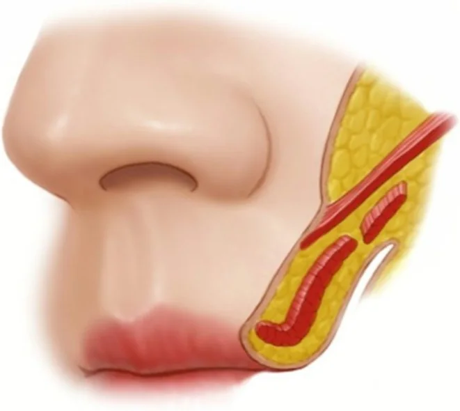
The youthful midface is a continuous smooth convexity from the lower eyelid to the nasolabial fold, with a short lid-cheek junction with no visible transition. The first thing lost is that continuity: a depression appears at the junction between the thin eyelid skin and the thicker cheek skin, medially first and laterally in time. In parallel, fat redistributes — accumulating anteriorly and inferiorly, lost superiorly and laterally — and the malar convexity flattens or inverts. Three findings follow, examined together: the nasojugal or tear trough, running two to three centimetres inferolaterally; the submalar depression, an inverted triangle bounded by the zygoma above, the nasolabial fold medially and the masseter laterally; and preauricular hollowing from loss of lateral fat.
What to look at on examination
- Examine at rest and smiling: the midface changes completely with animation and many defects only appear then.
- Assess surface contour and shadows, dermal and fat pad atrophy, dynamic and static lines, and loss of malar projection and bone volume.
- Locate the point of maximal malar projection: ideally 10 mm lateral and 15 mm inferior to the lateral canthus.
- Mark the infraorbital foramen before touching anything. It lies 6-8 mm below the arcus marginalis.
The vascular map to keep in mind
The infraorbital artery emerges from its foramen, 6 to 8 mm below the rim, supplying the lower eyelid, lateral nose and upper lip; it anastomoses with the transverse facial, angular, buccal and with branches of the ophthalmic and facial arteries. In the nose, supply comes from the facial, with two branches that matter: the lateral nasal and the dorsal nasal. And here lies the critical point of the whole face: the dorsal nasal anastomoses with the supratrochlear, meaning the external carotid territory communicates with the internal. An embolus injected under pressure there has an open route to the retina. This is not a theoretical risk nor one avoidable by dexterity: it is a consequence of anatomy, and it is why the nose is territory for experienced injectors.
Cheek and lid-cheek junction: where, how much and what to avoid
| Area | Plane and volume | What to avoid |
|---|---|---|
| Lateral cheek | Small supraperiosteal bolus. 0.1-0.3 ml per site, total 0.3-0.5 ml per side; if 0.5 ml is needed, split into two boluses | Zygomaticofacial vessels and nerves. Block with a finger so product does not migrate to the temple |
| Anterior cheek | Small deep subcutaneous bolus, or supraperiosteal if bony projection is lacking. Up to 0.3 ml per site, 0.5 ml per side | Infraorbital artery and vein. Always inject below the rim with a finger protecting the eyelid |
| Medial cheek | Supraperiosteal or deep subcutaneous, always lateral to the mid-pupillary line. Up to 0.3 ml per site | Angular and infraorbital arteries. Medial to that line and near the nose, a 25 G blunt microcannula and not a needle |
| Lid-cheek junction | Very small supraperiosteal boluses at two or three sites per region. Up to 0.1 ml per site and 0.5-0.6 ml per side | Experienced injectors only. Enter 1-2 mm below the rim, eyes closed. Risk of persistent eyelid oedema, embolisation, lumps and diplopia |
| Submalar | Subcutaneous: four boluses per side, or a single medial point with a fanning technique. Up to 0.25 ml per site, maximum 1 ml per side | Facial artery and vein, parotid duct and buccal branches of the facial nerve. Pinch the skin, aspirate, and massage intraorally as well |
| Preauricular | Small superficial subcutaneous boluses at three to five sites per side. 0.1-0.2 ml per site, up to 1 ml per side | Parotid and transverse facial artery and vein: the subcutaneous plane is mandatory. Prone to irregularity: small bolus, massage, repeat |
Two errors of judgement, not technique
First: overvolumising the cheek to erase the nasolabial fold. It does not work and leaves an artificial cheek that betrays itself on smiling; the fold is treated directly. The check is simple: volume must look natural at rest and in animation, and many results that convince in a resting photograph do not survive a smile. Second: fully correcting with a highly hydrophilic product at the lid-cheek junction. There it is wise to stop at around 50 % correction if the product attracts a lot of water, because late oedema appears weeks later and is among the hardest things to resolve.
The nose: four points and one common rule
| Goal | Where and how | Volume and caution |
|---|---|---|
| Frontonasal angle | A single supraperiosteal midline bolus, touching bone. Pinch the skin and hold the fingers so it does not spread laterally | 0.1-0.2 ml, and 0.2-0.3 ml in Asian patients. Beware the subcutaneous periorbital anastomoses |
| Bony dorsum | A single supraperiosteal injection, retrograde linear or as a small bolus | 0.1-0.2 ml. Avoid the dorsal nasal artery and vein |
| Cartilaginous dorsum | A single injection above the cartilage, retrograde linear | 0.1-0.2 ml. Avoid the dorsal nasal, external nasal, the external nasal nerve and the lateral nasal artery in the alar groove. Beware of deforming the supratip |
| Nasolabial angle and columella | Supraperiosteal bolus at the anterior nasal spine for the angle; retrograde linear in the anterior cartilaginous septum for the columella | 0.1-0.3 ml and 0.1-0.2 ml respectively. Do not inject into septal cartilage, avoid the columellar branches of the superior labial artery, and take care not to lengthen the upper lip or widen the columella |
The rule common to the whole nose
It is always the same and worth automating: pinch the skin, touch bone or cartilage, aspirate, stay in the midline, and inject very slowly at low pressure. Pressure matters as much as depth: an embolus occurs when local arterial pressure is exceeded, and that depends on injection speed. And a stopping rule that admits no nuance: on any change in skin colour or intense pain, stop the injection at that moment. Two further warnings. In patients with previous nasal surgery complication risk rises substantially and the reasonable course is not to treat them, or to let their surgeon do it. And in very flat noses the blunt microcannula helps but does not eliminate the risk of blindness or necrosis.
The little that toxin does here
| Indication | Target and technique | Dose |
|---|---|---|
| Bunny lines | Transverse nasalis, two lateral points and in some patients a third medial one. Needle to one third of its depth | 2 U per site, total 4-6 U |
| Nasal tip elevation | Depressor septi nasi, at the columellar base | 2-4 U |
| Nose widening on smiling | Corrected with the bunny line points plus one point at the columellar base: levator anguli oris is responsible | Around 2.5 U |
The bunny lines that appear afterwards
An effect worth anticipating in clinic rather than discovering at review. In some patients bunny lines did not exist before treatment and appear afterwards, following glabellar or crow’s feet injection, if nasalis was not also blocked: the muscle takes on the work its neighbours no longer do. It is not a technical failure but compensation, and it is resolved by adding the two transverse nasalis points. Warning about it beforehand turns a complaint call into an expected review.
A marked nasolabial fold: where does it come from?
Is it mainly marked on smiling?
Is there loss of malar projection above?
Why the cheek is treated first
- The midface is the supporting zone for the upper and lower thirds: what is placed here holds up what is above and below.
- Treating the lateral cheek indirectly improves the tear trough, nasolabial fold, commissure, marionette lines and jawline, and supports the brow.
- This is why a plan starting at the cheek needs less product elsewhere than one that goes area by area.
References
- 1.de Maio M, DeBoulle K, Swift A, Peng P, Weiner S. Facial assessment and injection guide for botulinum toxin and injectable hyaluronic acid fillers: focus on the midface. Plast Reconstr Surg. 2017;140(4):540e-550e.
- 2.Freilinger G, Gruber H, Happak W, Pechmann U. Surgical anatomy of the mimic muscle system and the facial nerve: importance for reconstructive and aesthetic surgery. Plast Reconstr Surg. 1987;80(5):686-690.
- 3.Mendelson BC, Muzaffar AR, Adams WP. Surgical anatomy of the midcheek and malar mounds. Plast Reconstr Surg. 2002;110(3):885-896.
- 4.Rohrich RJ, Pessa JE. The fat compartments of the face: anatomy and clinical implications for cosmetic surgery. Plast Reconstr Surg. 2007;119(7):2219-2227.
- 5.Khan TT, Colon-Acevedo B, Mettu P, DeLorenzi C, Woodward JA. An anatomical analysis of the supratrochlear artery: considerations in facial filler injections and preventing vision loss. Aesthet Surg J. 2017;37(2):203-208.
Related specialty: Facial Aesthetic Surgery