Principles of facial animation — Layers, innervation and why it matters when injecting
What sets a mimetic muscle apart, the four muscular planes and their three innervation exceptions, functional grouping, facial nerve territories, and what all this implies for palsy, toxin and filler.
Content intended for healthcare professionals. It does not replace clinical assessment and is not written as patient information.
Key points
- The mimetic muscles are the only ones in the body that insert into skin. That is why they move the face, and why their tone, atrophy and denervation are read directly on the surface.
- They are arranged in four planes, and the nerve reaches them on their deep surface — with three exceptions, which are precisely the three deepest muscles.
- That rule is what makes dissection safe: a plane kept superficial to layer 3 denervates nothing.
- A wrinkle lies perpendicular to the fibre direction of the muscle that produces it. This rule identifies the culprit muscle without seeing anything.
- The temporal and marginal branches leave permanent deficits, because they are the ones with fewest anastomoses with their neighbours.
What sets a mimetic muscle apart
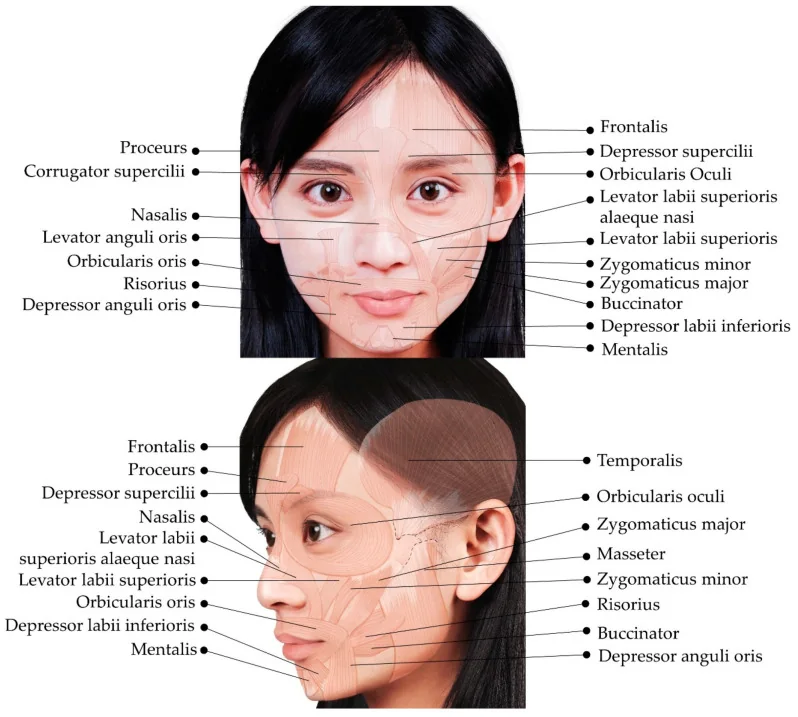
An ordinary skeletal muscle runs bone to bone and moves a joint. Mimetic muscles do not: they arise from bone or an aponeurosis and end in the dermis. That cutaneous insertion is what turns a contraction into an expression, and it has three consequences used daily. The first is that the wrinkle is the muscle’s footprint: it forms perpendicular to its fibre direction. The second is that there is no tendon to replace, so reanimation in palsy must reconstruct the vector, not only the muscle. And the third, subtler: mimetic muscle has no motor endplates gathered in a band as skeletal muscle does, but endplates spread throughout the belly, which explains why toxin works where you inject rather than at a single motor point.
Skeletal muscle and mimetic muscle
| Ordinary skeletal | Mimetic | |
|---|---|---|
| Distal insertion | Bone, via a tendon | Dermis, no tendon |
| What it moves | A joint | The skin: expression |
| Motor endplates | Grouped in a central band | Spread throughout the belly |
| Fibres | Larger, uniform calibre | Fine and highly variable calibre within the same muscle |
| Where the nerve enters | Variable | On the deep surface, with three exceptions |
The four muscular planes
The mimetic musculature is not a single sheet: it is organised in four superimposed planes, from superficial to deep. Knowing them answers two different questions. In surgery, it tells you what is being divided when a plane is opened. When injecting, it tells you at what depth to place the product to reach the muscle you want without diffusing into its neighbour, which often does exactly the opposite.
Which muscle is in each plane
| Plane | Muscles |
|---|---|
| 1. Most superficial | Orbicularis oculi, zygomaticus minor and depressor anguli oris |
| 2 | Zygomaticus major, levator labii superioris alaeque nasi, risorius, depressor labii inferioris and platysma |
| 3 | Orbicularis oris and levator labii superioris |
| 4. Deepest | Levator anguli oris, buccinator and mentalis |
The innervation rule and its three exceptions
The facial nerve approaches the mimetic muscles on their deep surface. This is the anatomical reason why any dissection kept superficial to them preserves animation intact, and why the subcutaneous plane is safe for the nerve even if it is poor for the skin. The exceptions are exactly three, and they are the three muscles of the deepest plane: levator anguli oris, buccinator and mentalis, which receive the nerve on their superficial surface. It makes sense: there is nothing deeper from which to approach them. The practical consequence is that in perioral and deep cheek dissection the protection the other layers give does not exist.
How to group them by function
The anatomical list is long and hard to retain; the functional list is short and used daily. Mimetic muscles do three things: elevate, depress and close or open an orifice. What matters is that almost all work in antagonist pairs on the same structure, so the visible expression is the result of a balance. When that balance breaks — through palsy, atrophy or a badly placed injection — what appears is not an absence of movement but the antagonist’s movement unopposed. This is the key to understanding both the paralysed face and the adverse effect of toxin.
The antagonist pairs
| Structure | Elevates or opens | Depresses or closes |
|---|---|---|
| Brow | Frontalis | Corrugator, procerus, depressor supercilii and the orbital orbicularis |
| Palpebral fissure | Levator palpebrae superioris (III nerve) and Müller’s muscle | Orbicularis oculi |
| Oral commissure | Zygomaticus major and minor, levator anguli oris, risorius | Depressor anguli oris and platysma |
| Upper lip | Levator labii superioris alaeque nasi, levator labii superioris | Orbicularis oris |
| Lower lip and chin | Mentalis, which elevates the chin | Depressor labii inferioris |
The rule that identifies the culprit
- The wrinkle lies perpendicular to the fibre direction. Look at which way the line runs to know which way the muscle runs.
- Horizontal forehead lines: vertical fibres, it is frontalis.
- Vertical glabellar lines: horizontal and oblique fibres, they are the corrugators.
- Horizontal line at the nasal root: vertical fibres, it is procerus.
- Crow’s feet: circular fibres, it is the orbital orbicularis.
- Perioral barcode lines: circular fibres, it is orbicularis oris.
Faced with a wrinkle: three questions
Is it present at rest or only on animation?
Is it a fold with volume loss beneath?
Is the problem the position of a structure, not the line?
The territories of the facial nerve
After traversing the parotid, the facial nerve divides into five branches: temporal — or frontotemporal — zygomatic, buccal, marginal mandibular and cervical. What orders surgical risk is not where each one runs, but how many connections it has with its neighbours. The zygomatic and buccal branches form a dense anastomotic network between them, so division of an isolated twig is usually compensated. The temporal and marginal branches, by contrast, are practically terminal: they have few connections, and injuring them produces a deficit that does not compensate itself. Hence the two classic permanent sequelae of facial surgery are the brow that will not rise and the lower lip that will not depress.
The five branches and what is lost with each
| Branch | What it supplies | What is lost if injured |
|---|---|---|
| Temporal | Frontalis, corrugator, depressor supercilii and the upper orbicularis | Dropped brow and smooth forehead. Few anastomoses: usually a permanent deficit |
| Zygomatic | Lower orbicularis and the lip elevators | Incomplete eyelid closure. Dense anastomotic network: usually compensates |
| Buccal | Buccinator, orbicularis oris and the upper lip elevators | Oral competence and smile. Also richly anastomosed |
| Marginal mandibular | Depressor anguli oris, depressor labii inferioris and mentalis | Lower lip asymmetry on smiling and speaking. Terminal branch: permanent deficit |
| Cervical | Platysma | Little functional impact; it can mimic a marginal injury |
How to tell a cervical injury from a marginal one
Both cause lower lip asymmetry and are easily confused. The difference lies in which movement is missing. If the injury is to the marginal branch, active eversion and depression of the lower lip fails — the patient cannot show the lower incisors — because the depressors are denervated. If it is to the cervical branch, the depressors work and what fails is the platysmal pull; the lip moves, though the gesture is less full. The clinical test is to ask the patient to show the lower teeth and then to tense the neck: if the lip descends but the platysma does not stand out, the injury is cervical, and its prognosis is far better.
Animation and the SMAS are the same layer
The superficial musculoaponeurotic system is not a structure separate from the mimetic muscles: it is the sheet that envelops and connects them. That is why orbicularis oculi and platysma are described interchangeably as mimetic muscles or as part of the SMAS, and why continuity runs uninterrupted from galea to neck. The consequence is twofold. In surgery, mobilising the SMAS means mobilising the whole mimetic apparatus, which explains why a well-performed facelift does not change expression: it moves the entire block without disconnecting anything. And in the operated face, that same continuity transmits tension from cheek to commissure, which is the mechanism of the marionette mouth when the vector is wrong.
What all this means in facial palsy
- The paralysed face is not a still face: it is a face pulled by the healthy side, because the antagonists on the affected side have stopped opposing.
- Hence weakening the healthy side with toxin improves symmetry without touching the paralysed side, and is often the first therapeutic step.
- The cutaneous insertion explains why reanimation must restore a vector, not merely supply working muscle.
- The diffuse endplate distribution is why denervation atrophy is fast and homogeneous throughout the muscle.
What it means when injecting toxin
- Inject into the belly that forms the wrinkle, not at a motor point: the endplates are spread out.
- The depth depends on the muscle’s plane: orbicularis is superficial and takes 2 mm; the corrugator at its medial insertion is deep.
- The adverse effect is almost always diffusion into an antagonist: hence the brow drops if frontalis is injected lateral to the mid-pupillary line.
- And conversely: blocking the depressor elevates. Treating the corrugator without touching frontalis opens the eyes better than any traction.
What it means when injecting filler
- Muscle is not merely an obstacle but a plane reference: knowing its layer tells you whether the product goes supraperiosteal, subcutaneous or intradermal.
- Product placed inside an active muscle migrates and resorbs sooner, besides distorting the expression.
- The dangerous vessels run in relation to specific muscular planes, so the muscular map and the vascular map are the same map.
- A fold with an overactive muscle beneath needs both: filler for volume and toxin so it does not re-etch.
The recurring mistake
Treating the line instead of the force that produces it. A wrinkle is the end result of a muscle pulling in one direction, over skin that has lost elasticity, over volume that has diminished. Filling the line without restraining the muscle guarantees its return; restraining the muscle without replacing volume leaves the depression; and treating the surface without doing either re-etches it within months. The useful question in clinic is not “what do I put in this wrinkle”, but “of the three causes, how much does each contribute in this particular face”.
References
- 1.Freilinger G, Gruber H, Happak W, Pechmann U. Surgical anatomy of the mimic muscle system and the facial nerve: importance for reconstructive and aesthetic surgery. Plast Reconstr Surg. 1987;80(5):686-690.
- 2.Happak W, Liu J, Burggasser G, Flowers A, Gruber H, Freilinger G. Human facial muscles: dimensions, motor endplate distribution, and presence of muscle fibers with multiple endplates. Anat Rec. 1997;249(2):276-284.
- 3.Freilinger G, Happak W, Burggasser G, Gruber H. Histochemical mapping and fiber size analysis of mimic muscles. Plast Reconstr Surg. 1990;86(3):422-428.
- 4.Davis RA, Anson BJ, Budinger JM, Kurth LE. Surgical anatomy of the facial nerve and parotid gland based upon a study of 350 cervicofacial halves. Surg Gynecol Obstet. 1956;102(4):385-412.
- 5.Mitz V, Peyronie M. The superficial musculo-aponeurotic system (SMAS) in the parotid and cheek area. Plast Reconstr Surg. 1976;58(1):80-88.
- 6.Pitanguy I, Ramos AS. The frontal branch of the facial nerve. Plast Reconstr Surg. 1966;38(4):352-356.
- 7.de Maio M, Swift A, Signorini M, Fagien S. Facial assessment and injection guide for botulinum toxin and injectable hyaluronic acid fillers: focus on the upper face. Plast Reconstr Surg. 2017;140(2):265e-276e.
Related specialty: Facial Paralysis