Fillers and vascular complication — Danger anatomy, prevention and what to do when it happens
Which territories link the face to the retina, which measures really reduce embolism risk, how an occlusion is recognised in its first minutes, and the high-dose pulsed hyaluronidase protocol.
Content intended for healthcare professionals. It does not replace clinical assessment and is not written as patient information.
Key points
- The face has open anastomoses between the external and internal carotid. That is why product injected in the cheek can reach the retina: it is not bad luck, it is anatomy.
- Embolism occurs when injection pressure exceeds local arterial pressure. That is why speed matters as much as depth.
- The most overlooked sign is immediate, disproportionate pain. Blanching, severe pain or visual change mean stopping at that instant.
- Treatment is not a dose: it is a protocol of high, repeated dosing until the tissue responds. The endpoint is clinical — colour, capillary refill and resolution of pain — not a number.
- With visual loss, the described useful window is about 90 minutes. That makes advance preparation — kit, phone number and an ophthalmology pathway — part of the treatment.
Two different mechanisms, two different timings
They are worth separating because they are not managed the same. Extrinsic compression occurs when excessive volume placed beside a vessel collapses it from outside; it appears progressively, over minutes or hours, and often improves with massage and volume reduction. Intravascular embolism is something else: product enters the lumen and travels, so the ischaemic territory does not match the injection point and the picture is immediate. The practical distinction is that compression gives you time and embolism does not. And there is a third, worst scenario: product injected under pressure into a facial artery can travel retrograde against flow until it reaches the ophthalmic system and from there embolise the retina or even the cerebral territory.
Which vessel, which area, and what is lost
| Vessel | Where it is injured | Consequence |
|---|---|---|
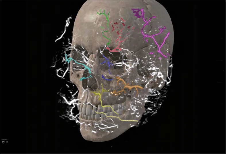
| Supratrochlear and supraorbital | Glabella and medial forehead, especially with superficial boluses | They are terminal branches of the ophthalmic: a direct route to the retina. Blindness and forehead skin necrosis |
| Dorsal nasal | Nasal dorsum and frontonasal angle | It anastomoses with the supratrochlear: it links external and internal carotid territories. Blindness and dorsal necrosis |
| Angular | Proximal nasolabial fold and medial cheek, close to the nose | Necrosis of the nasal ala and medial cheek, and through its continuity with the ophthalmic, visual risk |
| Lateral nasal | Alar groove | Nasal alar necrosis |
| Infraorbital | Anterior cheek and lid-cheek junction. It emerges 6-8 mm below the rim | Ischaemia of the lower eyelid and cheek; it anastomoses with the ophthalmic |
| Facial and labial | Lip and jawline. The labial arteries run in the submucosal plane, the wet part | Lip necrosis. It is where occlusion most often occurs, though not the most severe |
| Superficial temporal | Temple, in the subcutaneous plane | Scalp necrosis and alopecia. In the lower fossa there are also internal maxillary branches, with a risk of palatal necrosis |
The areas where it happens most
Reviews of the world literature on filler-induced blindness converge on a few sites: glabella, nose, nasolabial fold and forehead account for most published cases. This is no coincidence: they are exactly the areas where facial and ophthalmic territories communicate. It is useful information in clinic, because it orders the consent conversation: in the lip the complication is frequent but rarely catastrophic; in the glabella and nose it is infrequent but can cost an eye. It is worth saying it that way.
What does reduce the risk
- Know the planeBefore each area, review where the vessel runs and at what depth. It is the only measure acting on the cause rather than the consequence
- Inject slowlySlow, smooth and low pressure. A smaller gauge needle forces slower injection, and that is precisely what protects if the tip is inside a vessel
- Small volume per pointSplit into small boluses rather than one large one. The smaller the embolus, the smaller the ischaemic territory
- AspirateBefore each bolus. It does not guarantee being outside the vessel — a viscous product may not reflux — but it rules out many cases and takes two seconds
- Keep movingDeposit on withdrawal and while moving rather than static: it reduces the chance of unloading the whole volume into a lumen
- Cannula where appropriateIn areas of difficult anatomy, a blunt cannula of adequate gauge. It reduces vascular puncture, but does not eliminate it
- Block with a fingerPinching or compressing with the free hand occludes the proximal vessel and also prevents product displacement
What protects less than believed
Three beliefs worth qualifying. Aspiration is not a guarantee: with viscous products and fine needles there may be no reflux even with an intravascular tip, so a negative aspiration does not license fast injection. A cannula is not insurance: it lowers the incidence but occlusions and blindness have been described with cannulas, and in very flat noses or previously operated patients the risk persists. And experience does not immunise: published cases include expert injectors, because the mechanism does not depend on dexterity but on whether the tip is inside a lumen at the moment the plunger is pushed.
How it is recognised, and when
| Timing | Signs | What it means |
|---|---|---|
| During the injection | Intense, disproportionate pain, immediate blanching of the skin, or any ocular discomfort or visual change | Stop at that instant. Do not finish the point, do not "see if it improves" |
| Minutes | Persistent pallor, a reticulated or livedoid pattern, sluggish capillary refill, unremitting pain | Established occlusion. The affected territory indicates the vessel, which may be far from the injection point |
| Visual compromise | Vision loss, intense ocular pain, ptosis, ophthalmoplegia, headache, nausea or vomiting | Emergency. The described useful window is around 90 minutes: activate the ophthalmology pathway now |
| Hours to days | Violaceous discolouration, blisters, crust, and eventually an eschar | Established necrosis. No longer a dissolution problem but one of wound management |
| Neurological symptoms | Focal deficit, speech disturbance or altered consciousness | Cerebral embolism. Urgent transfer to hospital, not office management |
Pain is the sign, not the discomfort
Injecting is uncomfortable and every patient says so; what is not normal is intense, immediate pain, clearly disproportionate to what is being done. That pain is ischaemic and often precedes the colour change, particularly in thick or pigmented skin where blanching is hard to see. Two practical consequences. First, do not over-anaesthetise the area before injecting in risk regions: anaesthesia masks the only early warning there is. Second, ask actively during the compromised points — glabella, nose, nasolabial fold — rather than waiting for the patient to complain.
The first minutes
- 1. StopWithdraw the needle and do not inject one drop more in that area
- 2. AssessMap the blanched territory, check capillary refill and ask about vision and ocular pain. Vision is always asked about, even if the area seems remote
- 3. HyaluronidaseFlood the whole ischaemic territory, not just the injection point: the enzyme diffuses and crosses the vessel wall. Reconstitute with local anaesthetic without adrenaline if available: it hurts less and produces vasodilatation
- 4. Firm massageImmediately after each dose, vigorous massage to disperse the enzyme and break up the embolus. Local warmth to vasodilate
- 5. ReassessAt 15 to 20 minutes: if flow has not been restored, repeat the dose. The tissue half-life of the enzyme is minutes, not hours
- 6. EndpointStop when the tissue has regained colour, has good capillary refill and no longer hurts. Not when a number of units has been reached
The dose: high, repeated and clinically guided
- The high-dose pulsed protocol consists of flooding the ischaemic tissue block with enough enzyme to hydrolyse the product along its whole course, not injecting the entry point.
- Published figures give guidance: a minimum of around 450 to 500 units per ischaemic area, and up to 1,500 units across several cycles if more than one territory is involved.
- The practical recommendation in guidance is not to use concentrations below 1,500 units in 5 ml and to treat to effect, not to a fixed dose.
- The fear of "overdoing it" is miscalibrated: the enzyme also degrades native hyaluronic acid, but the body restores it within 15 to 20 hours.
- Ultrasound, when available, improves accuracy: it locates the deposit and confirms reperfusion.
Hyaluronidase allergy: what the evidence says
It is a frequent concern and is overstated in its severe form. Since 1949 very few published cases of allergic reaction have required adrenaline, and most of what is reported as "allergy" are local type IV reactions — oedema, erythema, urticaria at the site — appearing after an hour and resolving spontaneously. Severity does appear dose- and route-dependent: below 1,500 units at a local site the described reactions are local; with very high doses or intravenous administration generalised symptoms appear. There is also an effect confused with allergy that is not: concentrations above 1,500 units in 10 ml are irritant and produce erythema and swelling that settle within 24 hours. That is a pharmacologically predictable reaction, not an immunological one.
Who does need to be asked
- Bee or wasp sting allergy. Their venoms contain hyaluronidase, so there is a real risk of cross-reactivity.
- With a history of a large local reaction to a sting, estimated anaphylaxis risk is around 5 %; with a history of anaphylaxis to a sting, it may reach 60 %.
- If the patient has reacted to both bee and wasp, the shared allergen is more likely to be hyaluronidase itself.
- Also ask about drugs that make anaphylaxis harder to reverse: beta blockers and ACE inhibitors.
On intradermal testing
It is a widespread practice and its limits are worth knowing: no validated concentration exists for testing hyaluronidase allergy, and allergy guidelines recommend not using skin tests to screen for drug allergy in the absence of a compatible clinical history. A negative test at a non-validated concentration rules out nothing. And there is a dangerous paradox: precisely in patients with a history of hymenoptera anaphylaxis — the only ones in whom testing would make sense — doing it in the office is itself an anaphylaxis risk, and it should be done in an allergy unit with support. What remains genuinely useful is the clinical history: background, medication and any reaction on previous exposure.
Visual compromise: what is known and what is not
This is the scenario with the least margin and the weakest evidence, so precision matters. What is established: the window to reverse an ophthalmic artery occlusion before damage becomes irreversible is of the order of 90 minutes, and the obligatory course is immediate referral to ophthalmology, in parallel with treating the cutaneous territory. What is not established: the efficacy of retrobulbar hyaluronidase, described in isolated cases and still controversial — the enzyme diffuses poorly across the optic nerve sheath and published results are mixed. Presenting it as the treatment for filler-induced blindness would be inaccurate. The described adjuncts — ocular massage, lowering intraocular pressure, vasodilators, hyperbaric oxygen, antiplatelet therapy — have limited support and must not delay referral.
The skin has blanched: what to decide
Are there any visual or neurological symptoms?
Does the blanching resolve in seconds on withdrawing the needle and massaging?
And if the product is not hyaluronic acid?
What must be ready before injecting anyone
- Enough hyaluronidase for a full protocol, not one vial. An occlusion can consume more than a thousand units.
- Local anaesthetic without adrenaline to reconstitute it, and materials for massage and local warmth.
- Adrenaline and an anaphylaxis protocol: not because of hyaluronidase, whose risk is low, but because a drug is being given.
- An ophthalmology phone number that answers, agreed beforehand and not looked up on the spot. With a 90-minute window, the pathway is part of the treatment.
- And a consent form that names the complication: in risk zones the word blindness must be said, not "visual disturbance".
Two details that get forgotten
Do not inject hyaluronidase into an area treated with botulinum toxin in the previous 48 hours: the enzyme is a dispersal agent and will spread the toxin away from its target. And do not inject it over an area with active infection, unless biofilm is suspected and the patient is already covered with a broad-spectrum antibiotic. A third detail, for elective dissolution after a poor aesthetic result: it can be reassessed at 48 hours and repeated, but it is wise to wait at least two weeks before injecting filler again, until the swelling has settled, otherwise the result is unpredictable.
When necrosis is already established
If the patient presents late with established areas of necrosis, the problem has changed in nature: it is no longer about dissolving an embolus but about managing a wound. That means conventional wound care, cover against infection and optimal healing conditions, with the same criteria as for any skin loss. It is worth saying because there is a temptation to keep applying the emergency protocol when it no longer applies: a hyperbaric chamber alone is not adequate treatment for established necrosis. And once healed, the sequela enters the territory of scar revision, with its own timings and techniques.
The complications that are not vascular
- Early and self-limiting: erythema, oedema, pain and bruising. Warn about them beforehand; they need no treatment.
- Tyndall effect: bluish discolouration from product placed too superficially. It resolves with hyaluronidase.
- Delayed-onset nodules, weeks or months later: type IV hypersensitivity, granuloma or biofilm, often mixed. If the product is hyaluronic, there is the advantage of being able to dissolve the nidus; if an infective component is suspected, only under antibiotic cover.
- Poor aesthetic outcome from misplaced, excessive or migrated product. It is reversible, and that reversibility is hyaluronic acid’s main advantage over any other filler.
References
- 1.DeLorenzi C. New high dose pulsed hyaluronidase protocol for hyaluronic acid filler vascular adverse events. Aesthet Surg J. 2017;37(7):814-825.
- 2.Murray G, Convery C, Walker L, Davies E. Guideline for the safe use of hyaluronidase in aesthetic medicine, including modified high-dose protocol. J Clin Aesthet Dermatol. 2021;14(8):E69-E75.
- 3.Beleznay K, Carruthers JDA, Humphrey S, Jones D. Avoiding and treating blindness from fillers: a review of the world literature. Dermatol Surg. 2015;41(10):1097-1117.
- 4.Beleznay K, Carruthers JDA, Humphrey S, Carruthers A, Jones D. Update on avoiding and treating blindness from fillers: a recent review of the world literature. Aesthet Surg J. 2019;39(6):662-674.
- 5.Botha J, et al. Causes and management of sight threatening complications of dermal filler injections: a review. Clin Exp Ophthalmol. 2024;52(3):365-373.
- 6.Cotofana S, Lachman N. Arteries of the face and their relevance for minimally invasive facial procedures: an anatomical review. Plast Reconstr Surg. 2019;143(2):416-426.
- 7.Khan TT, Colon-Acevedo B, Mettu P, DeLorenzi C, Woodward JA. An anatomical analysis of the supratrochlear artery: considerations in facial filler injections and preventing vision loss. Aesthet Surg J. 2017;37(2):203-208.
- 8.de Maio M, DeBoulle K, Swift A, Peng P, Weiner S. Facial assessment and injection guide for botulinum toxin and injectable hyaluronic acid fillers: focus on the midface. Plast Reconstr Surg. 2017;140(4):540e-550e.
Related specialty: Facial Aesthetic Surgery