Lower third and neck musculature — The modiolus, mentalis and platysma
Orbicularis oris, depressors, mentalis, buccinator, masseter and platysma: where they lie, what they do, and the doses and depths that separate a good result from an asymmetric smile.
Content intended for healthcare professionals. It does not replace clinical assessment and is not written as patient information.
Key points
- All the perioral muscles converge on the modiolus, a fibromuscular knot lateral to the commissure where elevators and depressors pull in opposite directions from the same point.
- Here filler comes first and toxin after: structure and support are provided first, and only then is it assessed which dynamic line remains.
- The commonest error has a specific name: diffusion into depressor labii inferioris, leaving a very visible asymmetric smile for months.
- Two landmarks always to palpate before injecting the jawline: the facial artery and the marginal branch, which cross superficially just anterior to the masseter.
- In the neck the rule is superficial and low dose: injecting deep or loading the dose causes difficulty swallowing.
The muscles, one by one
| Muscle | From where to where, and in which layer | What it does |
|---|---|---|
| Orbicularis oris | A ring around the mouth formed largely by fibres of the muscles converging on it. Layer 3 | Closes and protrudes. Its superficial fibres are responsible for protrusion, and its circular fibres produce the perioral barcode lines |
| Depressor anguli oris | From the oblique line of the mandible to the modiolus, triangular. Layer 1, the most superficial | Lowers the commissure: it produces the sullen or dissatisfied expression and the crease descending from the corner of the mouth |
| Depressor labii inferioris | From the oblique line to the lower lip, medial and deep to depressor anguli oris. Layer 2 | Depresses and everts the lower lip. It is what shows the lower incisors on speaking, and its palsy is the classic marginal branch sequela |
| Mentalis | From the mandibular incisive fossa to the chin dermis. Layer 4, the deepest, with the nerve on its superficial surface | Elevates the chin and protrudes the lower lip. Its hypertonia gives the cobblestone chin and deepens the mentolabial crease |
| Buccinator | From the alveolar processes and pterygomandibular raphe to the modiolus. Layer 4, with the nerve on its superficial surface | Presses the cheek against the teeth: oral continence, chewing and blowing. In palsy it is why food pools in the vestibule |
| Platysma | From the pectoral and deltoid fascia to the mandible and modiolus. It is the continuation of the SMAS in the neck | Lowers the jaw and the commissures. Its contraction creates vertical bands and blurs the jawline |
| Masseter | Not a mimetic muscle: it is masticatory, innervated by the trigeminal. From the zygomatic arch to the mandibular angle | Elevates the mandible. Its hypertrophy from bruxism widens the lower third and is a frequent toxin indication |
The modiolus: where everything crosses
It is a fibromuscular knot lateral to the commissure, where elevators — zygomatics, levator anguli oris, risorius — and depressors — depressor anguli oris, platysma — converge. That is the explanation for almost everything that goes wrong here: muscles doing opposite things share one insertion point, so product placed with a margin of millimetres can block the wrong antagonist. It also explains why the commissure is so sensitive to the vector in surgery: any traction on the SMAS eventually reaches the modiolus, and if the vector is horizontal the commissure goes backwards instead of upwards.
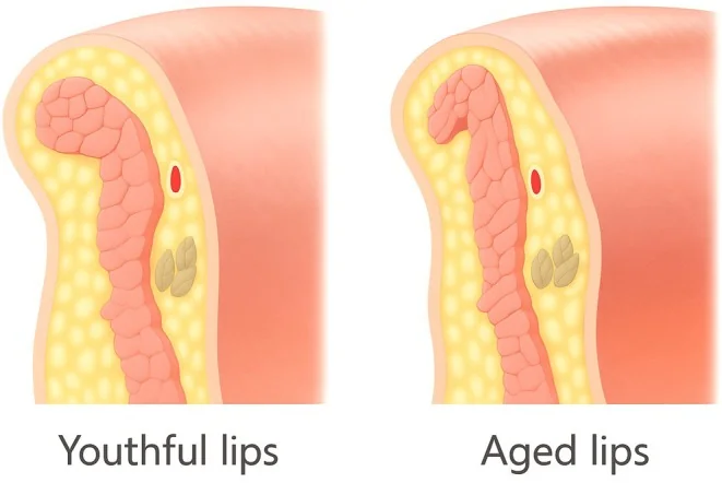
How the lower third ages
The youthful lip has a visible transition line between vermilion and skin, a V-shaped Cupid’s bow, a full medial tubercle, commissures with an ascending line, and an upper-to-lower lip ratio of 1 to 1.618. With age those elements are lost in a recognisable order: the vermilion flattens, the lip lengthens and thins, the Cupid’s bow blurs, the commissures turn downward and the mucosa dries. Around it, lines perpendicular to the lip border appear from perioral volume loss, lip atrophy, repeated muscular contraction and mandibular bone resorption beneath. The causal order matters: much of what looks like a lip problem is really loss of bony support and perioral volume.
What to examine before touching the lip
- At rest, smiling and puckering, always including lip projection on the profile view.
- Check what remains of the landmarks: vermilion border, body, Cupid’s bow, philtrum and commissures.
- Examine the dental arcade: occlusion and tooth inclination. A lip rests on what is behind it.
- Look for asymmetry at rest and in animation, excessive vermilion inversion and gingival show.
- And a sequence rule: before treating the nasolabial fold, the midface must have been assessed and treated.
Lip and perioral filler
| Area | Technique and plane | Volume and caution |
|---|---|---|
| Lip border | Entry at the border near the commissure, needle below the mucocutaneous junction, anterograde linear thread | 0.1-0.2 ml per quadrant. Equal volumes on both sides unless there is gross asymmetry. Do not overcorrect: it gives excessive anterior projection of the upper lip |
| Vermilion | Two options: linear thread in the dry submucosa with one point per quadrant, or aliquots at three points per quadrant. Needle at 30 degrees to the lip body | 0.5-1 ml for both lips, or 0.05 ml per bolus. Avoid the labial artery and vein in the intraoral submucosal plane, the wet part. Very bruise-prone: enter through skin and very slowly |
| Commissures | One point per side, very slowly | 0.05-0.1 ml per side. Avoid the labial artery and vein |
| Philtrum | Pinch the philtral column, enter at the base with the needle upward and the bevel inward, superficial subcutaneous retrograde | 0.05-0.1 ml per side. Preserve the inverted V of the philtrum, do not widen the columns and do not lengthen the upper lip. Avoid the columellar branches of the superior labial artery |
| Perioral lines | Needle perpendicular to the wrinkle, superficial subcutaneous linear, with massage after each point | Up to 0.25 ml per quadrant. Do not chase superficial lines: in many aged lips judicious volumisation improves the lines by itself |
Chin and jawline filler
| Area | Technique and plane | Volume and caution |
|---|---|---|
| Marionette lines | Two points per side. The lower one, retrograde linear superficial subcutaneous; the upper one, entering below the modiolus with a vertical column technique. Stay medial to the line | 0.5-1 ml per side at each of the two points. Deposit most of it in the upper third of the fold. Avoid the inferior labial and sublabial arteries |
| Mental crease | One point per side, retrograde linear superficial subcutaneous, with massage afterwards | 0.2-0.5 ml per side. Avoid the sublabial artery and vein. Overcorrection here leaves irregularities |
| Chin apex | Two or three supraperiosteal boluses: one midline and two superolateral. Pinch the chin with two fingers and aspirate first | 0.2-0.3 ml per site. Avoid the mental artery and vein. Do not inject too low: it produces a witch’s chin. Check symmetry from the cephalic view |
| Prejowl area | Triangular area between the mental foramen and the midlateral mandible | 0.5-1 ml per side. Avoid the mental artery, vein and nerve. Take care not to displace product over the mandibular ligament: overcorrecting lateral to it worsens the jowl |
| Mandible body and angle | Body: pinch the skin and inject superficially, retrograde linear, without going deep. Angle: one or two supraperiosteal boluses | 0.5-1 ml subcutaneous and 0.25 ml per bolus. The supraperiosteal angle injection is ideal in men; in women subcutaneous is preferred. Palpate and avoid the facial artery, facial vein and parotid. A zone of deep haematomas |
Toxin in the lower third and neck
| Indication | Points and depth | Dose |
|---|---|---|
| Gummy smile | Three points if moderate — levator labii superioris alaeque nasi and depressor septi — five if severe, adding levator labii superioris and zygomaticus minor. Needle at half depth, angled upward | 2 U per site. Total 6 U if moderate, 10 U if severe |
| Lip lines | One or two points per side on the upper, one per side on the lower. Insert only the bevel, bevel facing upward | 1 U per site. Total 2-4 U upper lip and 2 U lower |
| Depressor anguli oris | One point per side, near the jawline and more than 1 cm from the corner of the mouth, at half depth | 2-4 U per site, total 4-8 U |
| Mentalis | Midline point 0.5-1 cm above the lowest point of the chin and no closer than 1.5 cm to the lip. Needle at full depth, in the midline | 4-8 U. Two lateral points parallel to the midline may be added |
| Masseter | Three points per side, needle perpendicular and full depth, always lateral to the anterior margin of the muscle. Mark with the patient clenching | 4-8 U per site, total 4-24 U. Severe hypertrophy may need 40 U or more |
| Platysma at the jawline | Six points per side, needle at one third depth | 2-4 U per site, total 12-24 U per side |
| Platysmal bands | Four points per lateral band and three per medial band. Pinch the band to guide the needle into the contracted muscle, at one third depth | 2 U per site: 8 U per lateral band and 6 U per medial band |
The three errors that produce an asymmetric smile
All three share one cause: diffusion into depressor labii inferioris, which lies medial and deep to depressor anguli oris. One: injecting depressor anguli oris too medially or with too high a dose. Two: displacing the needle laterally when treating mentalis instead of staying in the midline. Three: injecting the lip too close to the mouth, which can also impair function and cause drooling. In all three the result is the same and lasts as long as the toxin. Prevention is geometric: 1 cm of margin from the commissure for depressor anguli oris, 1.5 cm from the lip for mentalis, and strict midline. And one habit: always review at two weeks, because this is the area with the highest risk of an asymmetric result.
The neck: superficial, and low dose
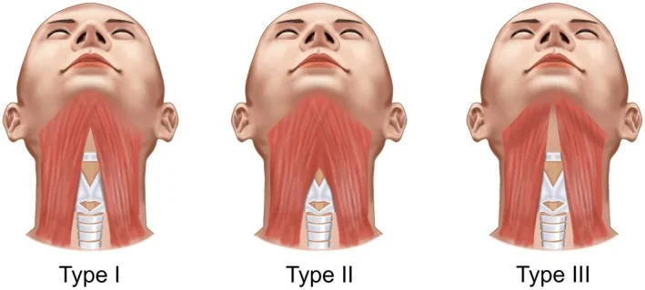
The complication to avoid here is not aesthetic: injecting deep or with excessive dose impairs swallowing. The rule is one third of the needle and a contained dose. Two further nuances. Start with the lateral bands and reassess at a second session whether the medial ones need treating: the medial bands are technically harder and their total dose should be lower. And select the patient carefully: reducing medial band hypertonia can leave skin laxity, so the ideal indication is a patient without skin excess in that area. In patients with a very active platysma, two sessions to titrate the dose give a better result than one loaded session.
The microdroplet technique
This is a different resource from muscular points, and it addresses what those do not: the quality and contour of the skin of the lower third and neck. It consists of depositing product in the dermis or at the junction between dermis and the superficial platysmal fibres — which insert precisely into the deep surface of the dermis — over an area beginning three fingerbreadths above the mandibular border, one fingerbreadth behind the marionette line, and extending across the whole neck anterior to the sternocleidomastoids. The described result is improvement of the cervicomental angle, elevation and flattening of the jowls, reduction of horizontal creases and vertical banding, and better skin texture. And there is a safety advantage: by distributing microdroplets in a superficial plane, the risk of dysphagia and sternocleidomastoid weakness is reduced.
Where the facial artery and marginal branch cross
Both cross superficially just anterior to the masseter, in the postjowl sulcus. It is the point to palpate before touching the jawline, and it explains two rules: in the mandibular body one does not inject deep, but pinching the skin in a superficial plane to stay above the artery; and at the angle, where the bolus is supraperiosteal, one must aspirate and expect deep haematoma. It is also worth remembering where the nerves exit: the mental through its foramen below the second lower premolar, and the infraorbital 6-8 mm below the orbital rim. Upper lip sensation depends on the infraorbital and lower lip on the mental; motor supply to both comes from the buccal branch.
Downturned commissure: three causes, three answers
Does it worsen on animation or is it the same at rest?
Is there an established marionette fold with volume loss?
Order matters
- In the lower third filler weighs more than toxin, the reverse of the upper third: structure and support first, dynamic lines after.
- Before the nasolabial fold, the midface. Before the commissure, the jawline. Before the lip, the dental arcade.
- Assess frontally and in profile: chin and jawline are only judged properly in profile, and the upper lip to lower lip and chin proportion should be one third to two thirds.
- A chin-neck angle of 121 degrees is considered optimal in women, and the jawline should run smooth from angle to chin, uninterrupted by jowl or sulci.
References
- 1.de Maio M, Wu WTL, Goodman GJ, Monheit G. Facial assessment and injection guide for botulinum toxin and injectable hyaluronic acid fillers: focus on the lower face. Plast Reconstr Surg. 2017;140(3):393e-404e.
- 2.Freilinger G, Gruber H, Happak W, Pechmann U. Surgical anatomy of the mimic muscle system and the facial nerve: importance for reconstructive and aesthetic surgery. Plast Reconstr Surg. 1987;80(5):686-690.
- 3.Ellenbogen R, Karlin JV. Visual criteria for success in restoring the youthful neck. Plast Reconstr Surg. 1980;66(6):826-837.
- 4.Furnas DW. The retaining ligaments of the cheek. Plast Reconstr Surg. 1989;83(1):11-16.
Related specialty: Facial Aesthetic Surgery