Botulinum toxin in facial palsy — Symmetrising by weakening, not stimulating
Why weakening the healthy side improves the paralysed face, how to assess before injecting, which muscle to treat on each side at what dose and depth, and what EMG or ultrasound guidance adds.
Content intended for healthcare professionals. It does not replace clinical assessment and is not written as patient information.
Key points
- The paralysed face is not a still face: it is an unbalanced one. Hence the most effective treatment is usually to weaken the healthy side, not to try to move the affected one.
- There are three distinct problems requiring three distinct answers: static and dynamic asymmetry, hyperkinesis of the healthy side and synkinesis of the affected side.
- The golden rule of the plan: start below usual doses and retouch at two weeks. Overtreating here is not a minor aesthetic effect but a temporary worsening of an already asymmetric face.
- The benefit outlives the pharmacology: combined with neuromuscular retraining, improvement persists beyond the toxin’s 3 to 6 months of effect.
- It is reversible, which makes it the best trial before reanimation surgery: it lets the patient rehearse the result before deciding.
The reasoning: why weakening works
Facial animation is a balance between antagonist pairs. When one side stops pulling, the other keeps pulling the same, and what is seen is not an immobile hemiface but a face dragged towards the healthy side, both at rest and on animation. Toxin produces a reversible chemical denervation blocking presynaptic acetylcholine release for 3 to 6 months, and that allows something no other tool offers: selectively weakening what is in excess without touching the little that works. It is applied in two different directions, and they should not be conflated: on the healthy side, against hyperkinesis; on the affected side, against the synkinesis of aberrant reinnervation.
Three problems, three targets
| Problem | What is seen | Where to inject |
|---|---|---|
| Asymmetry at rest | Brow, nasolabial fold and commissure at different heights without any expression | Healthy side: frontalis and depressors, to lower resting tone to the level of the affected side |
| Contralateral hyperkinesis | On smiling the mouth deviates markedly towards the healthy side, showing too much of its teeth | Healthy side: zygomatics, lip elevators, risorius, depressor anguli oris and depressor labii inferioris |
| Synkinesis | The eye closes on smiling, or the mouth moves on eye closure. It is aberrant reinnervation | Affected side: orbicularis oculi, platysma, depressor anguli oris and mentalis are the commonest targets |
Acute and chronic: where the cut-off lies and why it is debated
The classic division places the boundary at 18 to 24 months from onset, and it remains useful for orientation. But it is a temporal criterion applied to a biological problem, which is why it is being replaced by a better one: electromyography, which tells whether muscle remains viable by the presence of fibrillations. A muscle that fibrillates retains reinnervation potential, whatever the elapsed time; one that does not, does not. That distinction changes the goal of treatment: while viable muscle remains, the priority is protecting the recovery window; once it does not, the priority becomes symmetry.
Treating the healthy side may accelerate recovery of the affected one
It is the least intuitive and most useful finding. In the acute phase, the classic indication for toxin was purely symmetrising: reducing aesthetic and social impact while the nerve recovers. But several rehabilitation studies point to something more: reducing overactivity of the healthy side improves and accelerates motor recovery of the paralysed side, by lowering inhibitory feedback and facilitating neuromuscular retraining. Along the same lines, chemical denervation has been proposed to induce functional reorganisation of cortical networks, which would explain why improvement persists once the drug is gone. With the available evidence this is best presented as a well-founded hypothesis rather than an established fact, but it is reason enough not to delay treatment as a matter of routine.
The window of opportunity
The concept organises the whole treatment. While toxin keeps the abnormal neuromuscular transmission interrupted, simultaneous involuntary contractions disappear, and during that period the patient can train a correct movement pattern that was previously impossible because the intended gesture and the parasitic one came out together. That training — biofeedback exercises, work in front of a half-mirror — is what turns a months-long pharmacological effect into lasting improvement. Put differently: the toxin opens the window and rehabilitation is what goes through it. Injecting without pairing retraining wastes half the treatment.
Assessment before injecting
- HistoryCause, date of onset, course, previous treatments and whether there was partial recovery. Aberrant reinnervation only appears if recovery occurred
- Resting toneAssess the face globally and at rest before asking for any expression: this is where the static asymmetry corrected by baseline dosing shows
- Battery of expressionsA sequence that is always the same: raise brows, frown, gentle and forced eye closure, wrinkle the nose, small smile, broad toothy smile, pucker, whistle, and depress the lower lip
- ProfileAdd a profile view with platysmal contraction: it is the most forgotten muscle and the one with the highest yield
- DocumentPhotograph and video of the whole sequence at every visit. Video captures synkinesis far better than stills, because it is a movement phenomenon
- ScoreApply always the same scale and record it. Without a baseline score there is no way to know whether the next session improved anything
Which scale to use
| Scale | What it measures | When to use it |
|---|---|---|
| House-Brackmann | Six grades, from normal to total palsy. Global assessment: appearance, resting symmetry and movement of forehead, eye and mouth | Rapid grading in general clinic and emergency. It misses small changes and does not score synkinesis separately |
| Sunnybrook | Composite 0 to 100 score with three dimensions: resting symmetry, voluntary movement excursion across five expressions, and synkinesis | The reference in a dedicated clinic: far more sensitive to change and it allows each third to be assessed separately |
| Synkinesis questionnaire | The synkinesis burden as reported by the patient | It complements the others, which capture nothing the patient perceives |
The limits of the scales
Worth knowing so as not to over-read a number. House-Brackmann is simple but does not detect small changes nor separate synkinesis, which is precisely what is being treated. Sunnybrook solves that, but its interobserver reliability is only moderate, and the subscore that varies most between observers is precisely synkinesis. Neither includes patient-reported measures, so improvement in quality of life and social interaction goes unrecorded. Practically: pick one scale and apply it identically every time, add a synkinesis questionnaire, and do not compare scores taken by different observers.
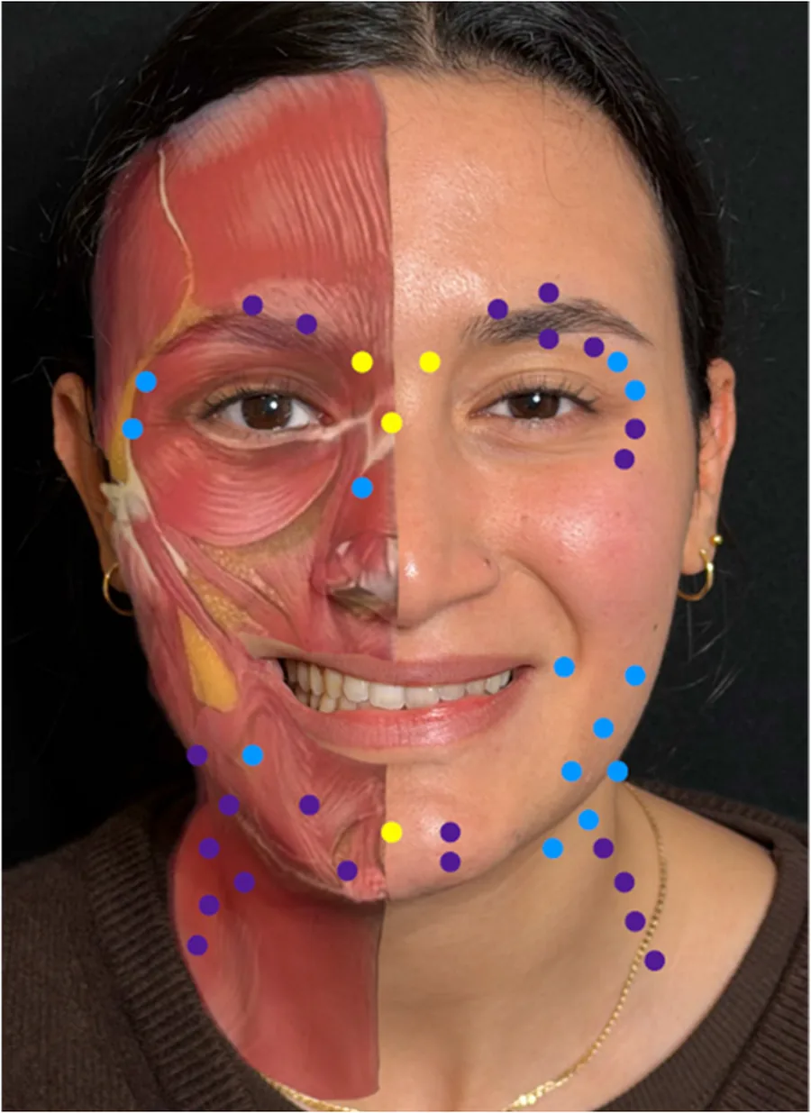
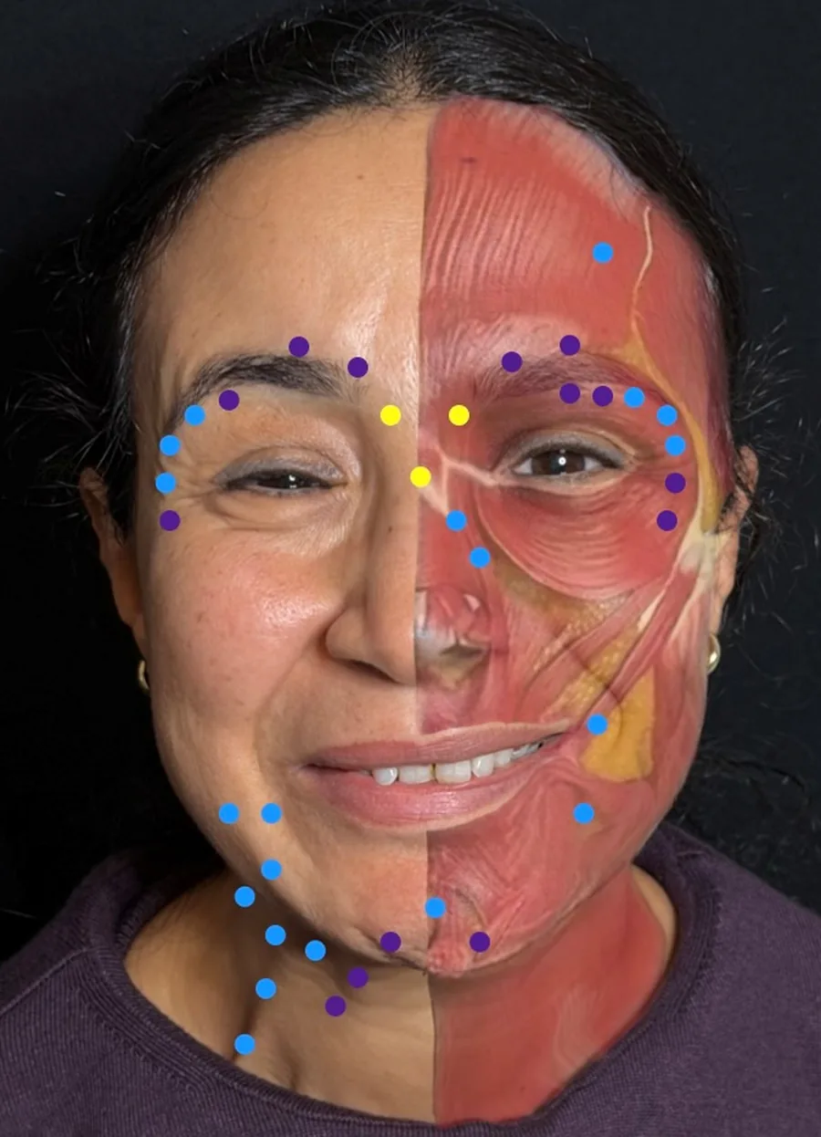
Affected side: upper third and orbit
| Muscle | Dose per session | Plane and depth |
|---|---|---|
| Procerus | 4-8 U | Intramuscular, 6-8 mm |
| Corrugator | 4-10 U | Intramuscular, 6-8 mm at the origin and 2 mm at the insertion |
| Depressor supercilii | 1-2 U | Subdermal, superficial |
| Frontalis | 1.5-18 U, the widest range of all | Intramuscular or intracutaneous, 2-4 mm |
| Orbicularis oculi | 2.5-10 U | Intracutaneous, 2-3 mm |
| Orbicularis, endopalpebral portion | 2-4 U | Intracutaneous, very superficial, 1 mm |
| Nasalis, transverse portion | 2-4 U | Intramuscular, 3-4 mm |
Affected side: middle third, lower third and neck
| Muscle | Dose per session | Plane and depth |
|---|---|---|
| Zygomaticus major | 1-5 U | Intramuscular at the muscle origin, 8-10 mm |
| Zygomaticus minor | 2 U | Superficial intramuscular, 5 mm |
| Risorius | 1-3 U | Intramuscular, 4 mm |
| Levator labii superioris alaeque nasi | 2-7.5 U | Intramuscular, 4-8 mm |
| Levator labii superioris | 1-2 U | Intramuscular, 4-8 mm |
| Buccinator | 2-20 U | Endobuccal route, perpendicular to the mucosa |
| Orbicularis oris | 1-6 U | Superficial or intradermal, 1-2 mm |
| Depressor anguli oris | 0.5-10 U | Deep intramuscular at the bony origin, at 1 to 3 points along its line |
| Depressor labii inferioris | 1-3 U | Intramuscular, 2-3 mm |
| Mentalis | 2.5-10 U | Deep intramuscular, 6-10 mm |
| Platysma, lateral and anterior portions | 10-60 U: by far the muscle consuming the most units | Intracutaneous or superficial intramuscular, 1-2 mm |
Healthy side: where and how much
| Muscle | Dose per session | Plane and depth |
|---|---|---|
| Corrugator | 2-4 U | Intramuscular, 6-8 mm at origin and 2 mm at insertion |
| Frontalis | 2-10 U | Intramuscular or intracutaneous, 2-4 mm |
| Orbicularis oculi | 4-10 U | Intracutaneous, 2-3 mm |
| Levator labii superioris alaeque nasi | 2.5-7.5 U | Intramuscular, 4-8 mm. To reduce upper tooth show |
| Depressor labii inferioris | 2.5-5 U | Intramuscular, 2-3 mm. To reduce lower tooth show |
| Platysma | 20-34 U | Intracutaneous, 1-2 mm |
| Mentalis | 3-5 U | Intramuscular, 6-10 mm at origin and 2 mm at insertion |
How to read these doses
Three cautions before using them. First: they are practical guidance, not an evidence-based standard. No validated international guidelines exist for toxin in facial palsy; these ranges come from case series and published experience. Second: units are onabotulinumtoxin and ranges from other publications have been converted to that equivalence, so conversion must be redone for another formulation. Third: frontalis on the affected side has the widest range for a specific reason — 1.5 to 18 units: it depends on whether the patient shows flaccid hypotonia, in which case the healthy side is treated to match, or paradoxical hyperkinesis, in which case the affected side itself is treated.
The dosing strategy
- Start below the usual dose and review at two weeks to decide whether to add. It is the opposite of aesthetic practice.
- The first targets are usually two: orbicularis, for narrowing of the fissure on animation, and platysma with depressor anguli oris, for downward commissure deviation.
- Depressor anguli oris and platysma can be treated assertively, but built up gradually: reviewing every two weeks until that patient’s dose map is found.
- The aim is not perfect symmetry but the greatest symmetry achievable without freezing expression: an immobile symmetric face is not a good result.
- Use the session to also treat muscles with a purely aesthetic indication: it improves overall satisfaction at no added cost.
Instrumental guidance: when it pays off
Landmark-guided injection gives good results in expert hands and needs no equipment, but its accuracy falls for small or deep muscles. That is where the two available tools come in, contributing different things. Electromyography directly identifies which muscle portion contracts abnormally, which in synkinesis is exactly the question; it uses needles that record the signal and allow injection in the same manoeuvre. Ultrasound does not say what contracts but where the structures are in real time, and serves to avoid the diffusion responsible for ptosis, diplopia, lagophthalmos or dysphonia. In cadaver models, ultrasound guidance improves both localisation and delivered dose compared with landmark technique. What is reasonable in practice: landmarks with standardised maps and systematic retouch sessions as the baseline, reserving instrumental guidance for complex cases.
Complications and how to avoid them
The most relevant complication is iatrogenic asymmetry from over-weakening the treated muscle, and it is likelier the higher the initial dose: for some weeks the face may look worse than before. Perioral weakness causes transient difficulty drinking, eating, articulating or puckering, especially after injecting orbicularis oris, orbicularis oculi and platysma. And there are target-specific risks: treating oro-ocular synkinesis may worsen blepharoptosis, lagophthalmos and diplopia, and overtreatment can distort expression — a distorted smile, a dropped brow — or cause frank loss of function. The advantage over surgery is that all of this is self-limiting and resolves within the pharmacological window; the duty is to warn beforehand, because during those weeks the patient’s distress is real.
Toxin and reanimation surgery are not rivals
Toxin has three distinct roles around functional microsurgery, and they are worth separating. Before: as initial treatment and, above all, as a reversible simulation of the surgical result; the patient experiences what less synkinesis feels like and decides knowingly whether to go to theatre. During the process: in cross-facial nerve grafts with secondary microcoaptations, it reduces aberrant co-contractions and improves the environment in which the reinnervated muscle must learn to move. After: when motor function has returned but movements are uncoordinated during fibre regrowth, it serves to refine and correct that result. In already-operated patients, weakening contralateral hyperkinesis can also improve the function of the reanimated muscle, which stops working against an overactive antagonist.
The three formulations do not last the same
| Formulation | At 1-2 weeks | At 4 weeks |
|---|---|---|
| Onabotulinumtoxin | Improvement similar to the other two | 41 % improvement |
| Abobotulinumtoxin | Similar improvement | 42 % improvement |
| Incobotulinumtoxin | Similar improvement | 17 % improvement: it may need higher doses or shorter intervals |
What can and cannot be claimed
The evidence here is consistent in direction but weak in quality: retrospective series and small cohorts predominate, with very few randomised trials, heterogeneous outcome measures, short follow-up and incomplete adverse-event reporting. No official published guidelines exist. Three questions remain open: when to start, whether instrumental guidance beats landmarks, and what role formulation plays in duration of effect. It is honest to explain it to the patient in these terms: the treatment is reversible, safe in trained hands and with documented high satisfaction, but the dosing scheme is individualised and empirical, not a validated protocol.
What to expect, with figures
- The peak effect falls between 28 and 42 days, and the clinically relevant effect lasts on the order of 150 to 180 days.
- In patients already operated for reanimation, mean asymmetry reduction was 48.8 % at one month with 16.8 % remaining at six months.
- That late remainder included an 18 % rise in the paralysed side’s score: weakening the healthy side improves the reanimated side’s function.
- Patient satisfaction frequently persists beyond the 3 to 6 months of pharmacological effect, particularly if rehabilitation was combined.
References
- 1.Renga M, D’Emilio R, Salti G, Mogavero S, Papagni M, Biglioli F, Lozza A. Facial paralysis treatment and facial symmetrization with botulinum neurotoxin: a narrative review with illustrative clinical cases. Toxins. 2026;18(6):253.
- 2.House JW, Brackmann DE. Facial nerve grading system. Otolaryngol Head Neck Surg. 1985;93(2):146-147.
- 3.Ross BG, Fradet G, Nedzelski JM. Development of a sensitive clinical facial grading system. Otolaryngol Head Neck Surg. 1996;114(3):380-386.
- 4.Freilinger G, Gruber H, Happak W, Pechmann U. Surgical anatomy of the mimic muscle system and the facial nerve: importance for reconstructive and aesthetic surgery. Plast Reconstr Surg. 1987;80(5):686-690.
Related specialty: Facial Paralysis