Deep-plane facelift, step by step — From marking to closure
The complete deep-plane sequence: marking with the patient seated, incision design, subcutaneous undermining, SMAS elevation and ligamentous release, repositioning and closure.
Content intended for healthcare professionals. It does not replace clinical assessment and is not written as patient information.
Key points
- Marking is done with the patient seated, since the supine position displaces the tissue and falsifies the limits of dissection.
- The subcutaneous undermining is deliberately wide: the wider it is, the more tension the composite flap accepts without it falling on the skin.
- The ligaments are released in a specific order — orbicular and masseteric first, zygomatic last — because the first two open the space that makes release of the third safe.
- The rule that prevents most pareses: the correct plane is the one in which the nerve branches never come into view.
- The repositioning sequence is SMAS, platysma and lastly skin, and the skin is closed without tension: the whole load is borne by the deep plane.
Before starting
This note develops the execution of the deep plane and takes as settled the questions that precede it: the layer to be mobilised, the orientation of the vector and the adaptation of the cervical design to the submandibular gland, all addressed in Choosing the technique: planes and vectors. The anatomical references cited throughout the sequence — the ligament line, the deep spaces and the course of the facial nerve branches — are described in Fat compartments and retaining ligaments and in Nerves and vascular supply. For explaining what this technique adds over a classic facelift in consultation, the patient-facing article Deep plane facelift in Barcelona: what it adds over a classic facelift may help.
Preoperative marking with the patient seated
- Areas of volumetric augmentationMark the regions to receive fat: malar compartment, tear trough, prejowl, nasolabial sulcus and temporal region. The planes and the contraindicated territories are in Facial fat grafting
- Limits of the dissectionAnterior borders of both sternocleidomastoids and the cervical midline at the level of the thyroid cartilage
- Anatomical referencesLigament line, from lateral canthus to mandibular angle, and Pitanguy’s line, from the earlobe to 1.5 cm above the brow tail
- IncisionsSubmental incision and cervical midline, in addition to the S-shaped facial design. The cervical stage is developed in Platysmaplasty and cervical contour
The design of the incisions
The design differs between the sexes for an anatomical reason: the distribution of terminal hair. In men, an endaural incision carries beard hair towards the auditory canal, and the temporal extension blurs the sideburn contour unless it is drawn geometrically. The table sets out the design segment by segment.
Incision design by segment
| Segment | In women | In men |
|---|---|---|
| Preauricular | Endaural, with a geometric design at the intertragal notch | Preauricular anterior to the beard hair, so as not to displace it towards the canal |
| Cranial extension | Following the helix and the hairline | The same, with a geometric design in the temporal extension to preserve the sideburn contour |
| Caudal extension | Around the lobule and retroauricular region, following the concha rather than the sulcus | The same |
| Retroauricular | Through the triangular fossa to the occipital hairline, in an M | The same |
| Submental | 2 cm, placed 2 mm caudal to the submental crease | The same |
Two points about the incision
Do not bevel the blade near the hairline. The rationale is that after displacement skin of differing characteristics is apposed, requiring edge-to-edge contact without tension to heal imperceptibly; bevelling prevents that apposition. The second point concerns design: a transverse occipital incision within the scalp limits the amount of cervical skin that can be excised, and is therefore not recommended. The M-shaped retroauricular extension following the occipital hairline allows the excess to be removed without compromising the cervical result.
Subcutaneous undermining
- InfiltrationTumescent solution, with greater volume in the infraorbital region and the territory of the zygomatic ligament: it displaces the musculature against bone and facilitates identification of the plane
- SequenceBegin in the preauricular region and continue into the cervical region. Blade oriented towards the surface to remain supra-SMAS
- ExtentThe supra-SMAS dissection must connect the neck with the submental space, run along the entire mandibular border releasing the mandibular ligament, and reach the ligament line on the face
- RationaleThe deep-plane flap is composite: the wider the cutaneous undermining, the greater the tension the flap accepts without it falling on the skin
Preserving adipose tissue on the skin flap
When elevating the skin flap, a layer of adipose tissue must be preserved adherent to the dermis, without dissecting flush against it. Omitting this produces two consequences: darkening of the flap, through transparency of the underlying platysma, and contour irregularities manifesting months later. The same principle reduces the risk of necrosis, since a flap of sufficient thickness retains its subdermal plexus. For the same reason the tragal flap must remain redundant and its SMAS is removed preserving the hypodermal tissue intact, on which its vascular supply depends.
SMAS elevation and ligamentous release
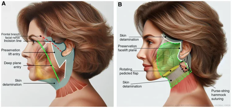
- Point of entrySMAS incision slightly posterior to the marked ligament line, drawing the SMAS-cutaneous complex superiorly to expose it
- Order of releaseRelease first the orbicular and masseteric ligaments, keeping the muscle in the deep plane in both, and the zygomatic last. After the first two, the prezygomatic space opens bluntly
- Dissection techniqueScissors held vertically, resting on the finger of the retracting hand and directing force towards the deep plane
- Anterior limitThe zygomaticus major and the facial artery delimit the anterior reach of the dissection
A clarification on the deep plane
The classical description places the deep plane immediately above the deep fascia. Recent anatomical work has qualified that definition: the plane used in practice is subplatysmal and deep subcutaneous, and may include part of the most superficial lamina of the deep fascia, so that the extent of the flap depends on the surgeon rather than on a strict anatomical boundary. From this follows an operative rule that prevents most pareses: the plane should not lie immediately on the fascia but somewhat more superficially, at a level where the nerve branches are not exposed. Their visualisation indicates that dissection has progressed too deeply.
Repositioning sequence: SMAS, platysma and skin
- Facial SMASSuspension with 3/0 absorbable monofilament to the fixed parotid SMAS, at 60 degrees, with a mattress stitch on the mobile SMAS to distribute tension. Five stitches, from the gonion to the temporal SMAS
- PlatysmaSuspension with 2/0 absorbable suture to the mastoid periosteum, at 80 degrees, likewise with a mattress stitch. Two points of fixation
- SkinSuperoposterior repositioning at around 60 degrees, close to the SMAS vector, followed by passive excision of the excess. The closure must bear no tension
- Order of closurePositioning stitch at the helical root → excision of the anterior excess → running suture in the temporal extension → stitch at the angle between concha and triangular fossa → excision of the posterior excess → cervical running suture → stitch at the lobule → excision of the remainder
- Residual excess at the endsFaced with a dog ear, extend the corresponding limb — temporal or cervical — rather than force the closure
Details of closure and the immediate postoperative period
- In the areas of greatest tension, simple absorbable stitches that fall spontaneously at 3-4 weeks: they maintain support through the critical period without leaving suture marks.
- Quilting stitches in the temporal and submandibular regions at the level of the gonion: they obliterate dead space, reduce the risk of haematoma and seroma, and are removed on the third day.
- The first dressing is applied without compression; compression is added from the first postoperative day, once the drains are removed.
- The full postoperative schedule and the management of complications are developed in Complications of facelift surgery.
Lines of technical evolution
- Cutaneous preservation: limiting skin dissection and extending the composite flap, rather than undermining widely in order to tension afterwards.
- Orbicularis revectorisation: incorporating the orbicularis into the mobile SMAS, extending the SMAS incision over the lateral orbicularis as a continuation of the flap.
- The manoeuvre includes release of the orbicularis retaining ligament, suborbicularis dissection to the premaxillary and prezygomatic spaces, release of herniated suborbital fat and superolateral resuspension of the orbicularis.
- Orbicularis paresis after these manoeuvres is exceptional, since the muscle receives innervation from three distinct branches — temporal, zygomatic and buccal — and that redundancy protects it.
References
- 1.Vaquero Martínez P. Cirugía de lifting. En: Manual de la Sociedad Española de Cirugía Oral y Maxilofacial y de Cabeza y Cuello. SECOM CyC; 2025.
- 2.Hamra ST. The deep-plane rhytidectomy. Plast Reconstr Surg. 1990;86(1):53-61.
- 3.Hamra ST. Composite rhytidectomy. Plast Reconstr Surg. 1992;90(1):1-13.
- 4.Jacono AA, Parikh SS. The minimal access deep plane extended vertical facelift. Aesthet Surg J. 2011;31(8):874-890.
- 5.Talei B. The detailed anatomy of the deep plane of the face and neck with current nomenclature. Facial Plast Surg. 2024;40(6):680-686.
- 6.Minelli L, Yang HM, van der Lei B, Mendelson B. Anatomy of the facial glideplanes, deep plane spaces, and ligaments: implications for surgical and non-surgical facial rejuvenation. Plast Reconstr Surg. 2023;154(1):95-110.
- 7.Pitanguy I, Ramos AS. The frontal branch of the facial nerve: the importance of its variations in face lifting. Plast Reconstr Surg. 1966;38(4):352-356.
- 8.Boyd CJ, Ceradini DJ. Current trends in facelift and necklift procedures. J Clin Med. 2025;14(12):4273.
Related specialty: Facial Lifting & Rejuvenation