Fat compartments and retaining ligaments — The anatomical origin of the facial sulci
The compartmentalisation of facial fat and the ligamentous system that delimits it, the mechanism by which both generate the sulci, the dissection line the ligaments describe and the recent reclassification of the mandibular ligament.
Content intended for healthcare professionals. It does not replace clinical assessment and is not written as patient information.
Key points
- Facial fat does not constitute a continuous layer but a set of compartments delimited by vascularised fibrous septa, which age independently of one another.
- The retaining ligaments fix the superficial layers to the deep plane. The septa separating the compartments are the superficial expression of that same system.
- The sulcus arises from the interaction of both: a compartment that descends and is arrested against a ligament that remains fixed. Every facial sulcus therefore has an identifiable compartment and ligament.
- The four ligaments that must be released align along a line running from the lateral canthus to the mandibular angle. That line defines where to release and, in aesthetic medicine, separates the territory of lifting effect from that of volumising effect.
- The mandibular ligament has been reclassified: it is not a true osteocutaneous ligament, and a specific surgical indication regarding its supraplatysmal portion follows from that.
Facial fat is compartmentalised
For a long time the subcutaneous fat of the face was regarded as a homogeneous mantle displaced en bloc with ageing. Injection and dissection studies have shown a different organisation: it is divided into independent compartments, separated by fibrous septa extending from the deep fascia to the dermis. Those septa are not inert partitions; they carry the perforating vessels that connect the deep plexus with the subdermal one, which explains why they constitute at once anatomical boundaries and axes of vascularisation.
Compartmentalisation has two consequences worth bearing in mind before describing each compartment. The first is that ageing does not affect them equally: some atrophy, others descend and others retain their volume, so that facial contour is altered by the loss of continuity between them rather than by a global descent. The second is surgical: raising the subcutaneous plane divides those septa and with them the perforators they carry, leaving flap vascularisation dependent on the supply remaining outside the undermined area.
The retaining ligaments
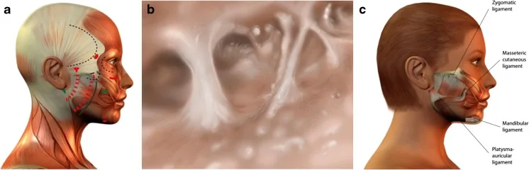
The retaining ligament system comprises the fibrous structures that fix the superficial layers of the face to the deep plane, preventing skin, hypodermis and SMAS from moving freely over the skeleton. Two types are distinguished by origin. Osteocutaneous ligaments arise directly from periosteum and reach the subcutis in an arborising fashion. Fasciocutaneous ligaments originate from the investing facial fascia rather than bone, and their fixation is less firm. The distinction matters because it determines what each produces: the former anchor discrete points and are the origin of the sulci; the latter create no sulci but hinder identification of the correct plane during dissection.
The ligaments, one by one
| Ligament | Type and location | Clinical relevance |
|---|---|---|
| Orbicularis retaining ligament | Osteocutaneous. From the orbital rim to the deep surface of the orbicularis muscle | Defines the lid-cheek junction. Its persistence maintains the palpebromalar sulcus and limits midface correction |
| Zygomatic | Osteocutaneous. From the malar body to the subcutis, with a dense contingent known as McGregor’s patch | Its release is indispensable to mobilise the midface. It is also the most predictable bleeding point of the dissection |
| Maxillary | Osteocutaneous. From the maxilla to the subcutis, at the medial limit of the nasolabial compartment | It is directly responsible for the nasolabial fold: it arrests the descent of the nasolabial compartment and marks its border |
| Mandibular | Classically osteocutaneous, now reclassified. Located in the anterior third of the mandibular body | It delimits the jowl anteriorly and contributes to the prejowl sulcus. Its true nature determines what should and should not be divided |
| Parotideomasseteric | Fasciocutaneous. Between the parotid and masseteric fascia and the subcutis | It generates no sulcus, but makes the plane harder to find where the facial nerve leaves glandular protection |
| Cervical (platysma-auricular) | Fasciocutaneous. Between the platysma and the auricular region | It retains the cervical angle. Its release is what allows facial traction to reach the neck |
How a sulcus forms
With both systems described, the mechanism of the sulcus can be stated precisely. The fat compartment descends and shifts medially; the ligament remains fixed; tissue accumulates above the anchoring point and thins below it. The depression the patient perceives is therefore neither a cutaneous rhytid nor an isolated volume loss, but the step formed between tissue that has moved and tissue that has not. The therapeutic consequence follows: filling the sulcus without releasing the ligament corrects the volume deficit but not the anchorage, and the step reappears with muscular movement.
Which compartment and which ligament lie behind each sulcus
| Sulcus | Compartments involved | Ligament arresting it |
|---|---|---|
| Nasolabial | Nasolabial compartment and the medial and middle cheek compartments | Maxillary |
| Melolabial | Nasolabial compartment and the upper portion of the jowl fat | Mandibular |
| Prejowl | Lower portion of the jowl fat, displaced anterior to the anchoring point | Mandibular |
| Palpebromalar and tear trough | Superficial malar compartments over the suborbicularis fat | Orbicularis retaining ligament |
The mandibular ligament, revised
Recent anatomical work has modified the classical description of this ligament, and the modification has an immediate surgical translation. It is not a true osteocutaneous ligament: it is formed not by fibres ascending from periosteum to dermis, but by a condensation of connective tissue at the insertion of depressor anguli oris and depressor labii inferioris onto the superficial surface of the platysma. Those fibres do not continue through the subcutis to reach the skin. The same revision established that the prejowl fat does not constitute a compartment of its own, but the continuation of the subcutaneous plane and of the hypodermal fat.
The consequence for technique
If the ligament does not reach the skin, dividing its supraplatysmal portion releases nothing and does destabilise the insertion of the depressors. The indication that follows is not to resect it. The prejowl sulcus, for its part, obeys two simultaneous mechanisms rather than one: a mechanical one, from accumulation of subcutaneous and hypodermal tissue above and platysmal tissue below, over a muscular plane firmly bound to bone in this region; and an atrophic one, from thinning of the superficial fat in a zone under permanent muscular traction, which also shadows the contour. Treating only one leaves the sulcus partly corrected. The approach covering both consists of filling with up to 1 ml of microfat in the subcutaneous plane — in the sulcus itself and, above all, in the indentation of the mandibular border — and reserving subcutaneous release of the ligament for cases where filling proves insufficient.
The ligaments describe a line
The four ligaments that must be released to mobilise the SMAS — orbicular, zygomatic, masseteric and cervical — are not randomly distributed over the face: they lie along a line running from the lateral canthus to the mandibular angle. That alignment has an immediate practical use, since it converts a set of anatomical points into a continuous reference: the ligament line is the release line, and marks how far the dissection must advance for the mobile segment to be genuinely free.
The same line, applied to filling
The usefulness of this reference extends beyond surgery. Since the line separates anchored from mobile territory, filler placed lateral to it acts on fixed tissue and produces a supporting effect; placed medial to it, it acts on mobile tissue and produces a volumising effect. This is the anatomical explanation of why two injections of equal volume give different results according to the entry point, and why the lifting effect sought in the midface depends more on where the material is deposited than on how much.
Why skin alone does not sustain the result
The anatomy described explains a well-established clinical observation. For decades facelift surgery consisted of raising the subcutaneous plane and repositioning the skin alone, and the result deteriorated predictably for two reasons: cutaneous elasticity returned the tissue to its initial position within months, and tension applied to the skin increased the risk of flap necrosis and hypertrophic scarring. The anatomical basis is that skin remains bound to the SMAS by the fibrous septa already described, so that pulling on it does not mobilise the plane that supports the contour. Dividing the ligaments is therefore not an optional step: it is what allows the mobile segment to be repositioned and, in doing so, relieves the skin envelope of tension.
References
- 1.Furnas DW. The retaining ligaments of the cheek. Plast Reconstr Surg. 1989;83(1):11-16.
- 2.Stuzin JM, Baker TJ, Gordon HL. The relationship of the superficial and deep facial fascias: relevance to rhytidectomy and aging. Plast Reconstr Surg. 1992;89(3):441-449.
- 3.Rohrich RJ, Pessa JE. The fat compartments of the face: anatomy and clinical implications for cosmetic surgery. Plast Reconstr Surg. 2007;119(7):2219-2227.
- 4.Rohrich RJ, Pessa JE. The retaining system of the face: histologic evaluation of the septal boundaries of the subcutaneous fat compartments. Plast Reconstr Surg. 2008;121(5):1804-1809.
- 5.Schaverien MV, Pessa JE, Rohrich RJ. Vascularized membranes determine the anatomical boundaries of the subcutaneous fat compartments. Plast Reconstr Surg. 2009;123(2):695-700.
- 6.Gierloff M, Stöhring C, Buder T, Gassling V, Açil Y, Wiltfang J. Aging changes of the midfacial fat compartments: a computed tomographic study. Plast Reconstr Surg. 2012;129(1):263-273.
- 7.Stuzin JM, Rohrich RJ, Dayan E. The facial fat compartments revisited: clinical relevance to subcutaneous dissection and facial deflation in face lift. Plast Reconstr Surg. 2019;144(5):1070-1078.
- 8.Mendelson BC, Jacobson SR. Surgical anatomy of the midcheek: facial layers, spaces, and the midcheek segments. Clin Plast Surg. 2008;35(3):395-404.
- 9.Seo YS, Song JK, Oh TS, Kwon SI, Tansatit T, Lee JH. Review of the nomenclature of the retaining ligaments of the cheek: frequently confused terminology. Arch Plast Surg. 2017;44(4):266-275.
Related specialty: Facial Lifting & Rejuvenation