Nerves and vascular supply — The structures that delimit the dissection
The course of the facial nerve branch by branch and its relation to the fasciae and ligaments, the safety principle that follows, the anatomy of the great auricular nerve and the vascular basis of the skin flap.
Content intended for healthcare professionals. It does not replace clinical assessment and is not written as patient information.
Key points
- The facial nerve leaves the parotid gland and runs within the thickness of the parotideomasseteric fascia above and the middle cervical fascia below. A dissection superficial to that fascia is safe for all its branches.
- Safety is resolved by planes rather than by direct vision: each branch has a plane in which it lies and another in which it cannot be, and that knowledge replaces systematic visual identification.
- The points of risk coincide with two anatomical events: where the nerve leaves glandular protection and where the fasciae fuse over the zygomatic arch.
- The great auricular nerve is the nerve most frequently injured in this surgery, and its sequela is sensory and permanent.
- The lateral skin flap depends on fasciocutaneous perforators that are divided during undermining, so that its distal portion is supplied only by peripheral inflow.
The principle that governs safety
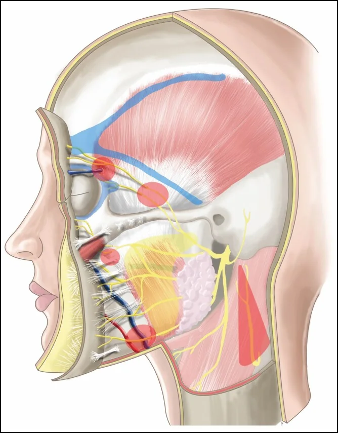
Protecting the facial nerve during facelift surgery rests not on identifying each branch but on knowing the plane in which it runs and remaining superficial to it. The basis is that, after leaving the parotid, the nerve does not travel free in areolar tissue: it is contained within the thickness of the deep fascia, termed the parotideomasseteric fascia in the upper territory and the middle cervical fascia below. From this follows an operative statement covering all four branches: any dissection remaining superficial to that fascia lies in a safe plane. The converse is equally relevant, and explains most injuries: seeking a plane within the fascia is not dissecting deeper, it is dissecting inside the compartment that contains the nerve.
The two points where the principle stops protecting
The rule above has two anatomical exceptions worth locating before operating. The first is the exit from the parotid gland: while the nerve runs beneath glandular parenchyma the gland acts as an interposition and the superficial plane is generous; anterior to its border that protection disappears and the margin narrows to the fascia itself. The second is the zygomatic arch, where the parotideomasseteric fascia fuses with periosteum: at that point the planes cease to be separable and the depth reference is lost. Both zones coincide with the most frequently reported sites of injury, and neither is identified by the depth reached but by position relative to those two landmarks.
The branches and their territory
The facial nerve divides within the gland into temporofacial and cervicofacial trunks, from which the terminal branches arise. Four cross the facelift field, and each has a specific motor function whose loss produces a recognisable deficit: the frontotemporal elevates the brow and wrinkles the forehead; the zygomatic closes the eyelid; the buccal moves the upper lip and nasal ala; and the marginal depresses the lower lip. Their relationship with the ligamentous system is not incidental: the branches cross the deep plane in the vicinity of the retaining ligaments, so that ligamentous release and the zones of risk share a location. The detailed study of each muscle’s innervation is developed in the section on the anatomy of facial animation.
Course of each branch and its safe plane
| Branch | Course | Safe plane |
|---|---|---|
| Frontotemporal | Follows Pitanguy’s line, crossing the zygomatic arch at its middle third | Two planes by level: below the arch, sub-SMAS and superficial to the parotid fascia; above it, deep to the deep temporal fascia. Over the arch both fasciae fuse and no separation plane exists |
| Zygomatic | Deep to the SMAS, within the masseteric fascia, and deep to the zygomatic musculature | Keep the zygomaticus major below the dissection plane. This is the reference that protects the branch during release of the zygomatic ligament |
| Buccal | Deep to the SMAS, within the masseteric fascia. It may become superficial to the buccal fat pad | Do not open the masseteric fascia. Manipulating the buccal fat pad demands care because the nerve may lie anterior to it |
| Marginal mandibular | Deep to the platysma, within the middle cervical fascia, until it becomes superficial at the depressor anguli oris | Do not dissect medial to the facial vessels: there the platysma separates poorly from the fascia and the nerve crosses superficial to the vein |
Sequence of entry to the deep plane
- General ruleDissect immediately deep to layer 3 — mimetic musculature, SMAS and platysma — and do not enter the thickness of the deep fascia
- FirstLower masseteric space, leaving the parotid capsule and masseteric fascia beneath. It offers the widest margin and serves as reference for the rest
- SecondSubplatysmal space, advancing towards the neck from the plane already identified
- ThirdPrezygomatic space, deep to the orbicularis, until the border of the zygomaticus major is exposed
- FourthZygomatic ligament, keeping the zygomaticus deep to the dissection to preserve the branch running beneath it
The great auricular nerve
The great auricular nerve arises from the superficial cervical plexus, from the C2 and C3 roots, and is purely sensory. That condition explains why it receives less attention than the motor branches, and contrasts with the fact that it is the nerve most frequently injured in facelift surgery. Its injury leaves an area of anaesthesia in the earlobe and retroauricular region that the patient notices daily and which, unlike motor pareses, does not usually recover.
Its course is well defined and admits reproducible references. It becomes superficial at Erb’s point, on the posterior border of the sternocleidomastoid, about 6.5 cm below the external auditory canal and roughly midway between the earlobe and the clavicle. From there it ascends obliquely over the muscle belly, one centimetre posterior and parallel to the external jugular vein, which is the most reliable intraoperative reference since it is visible through the fascia. It divides into three branches: posterior and lobular, both superficial and immediately subcutaneous, and anterior, deeper and intraparotid. As a whole it remains posterior to the platysma and adherent to the fascia investing the sternocleidomastoid, although its most superior and anterior branch may run within the SMAS itself.
How to preserve it
In cervical facelift, one measure protects it: raising the skin flap deliberately superficially over the sternocleidomastoid, leaving the nerve within the fascia investing the muscle. The manoeuvre is simple, and its difficulty is of another order: the area is approached once the dissection has become repetitive. Beyond facelift surgery, the same anatomy guides other procedures. In parotidectomy the platysma is opened at the anterior border of the sternocleidomastoid and the nerve can be dissected there, although the anterior branch is usually sacrificed with the specimen. In submandibular gland excision it is advisable not to dissect the platysma so cranially and to locate the nerve in the more posterior subplatysmal flap.
A clarification on the Hayes Martin manoeuvre
A frequent confusion is worth undoing. The Hayes Martin manoeuvre does not protect the great auricular nerve but the marginal mandibular branch of the facial nerve: it consists of ligating and dividing the facial vein below the mandibular border and reflecting it cranially, so that the marginal branch, which runs superficial to the vein, is displaced with it and away from the dissection field. It belongs to neck dissection and submandibular gland surgery, not to the approach to the great auricular nerve. Both structures are protected in the same region and are therefore easily conflated, but the plane and the manoeuvre differ: the great auricular is preserved by remaining superficial over the sternocleidomastoid; the marginal, by displacing the facial vein.
Vascular supply of the skin flap
The face receives a redundant arterial supply from the external carotid, with three main territories: the facial artery supplies the medial region, the superficial temporal the lateral and temporal region, and the posterior auricular and occipital the retroauricular and posterior cervical region. In the territory relevant to facelift surgery, the lateral cheek is supplied through fasciocutaneous perforators from the transverse facial artery, a branch of the superficial temporal, which ascend from the deep plane traversing the septa that delimit the fat compartments.
Why necrosis always appears in the same place
Subcutaneous undermining divides precisely those perforators, so that the flap comes to depend on inflow entering at its periphery. The most compromised portion is the distal part of the retroauricular flap, being furthest from any alternative source, and it is the site where necrosis is described when it occurs. The practical consequence is that the extent of undermining has a vascular limit as well as a technical one. A qualification about smoking is worth adding, since it is often stated imprecisely: the chronic smoker does not have fewer vessels but greater collateralisation from sustained hypoxia; the problem is not vascular density but effective perfusion and impaired tissue repair.
References
- 1.Pitanguy I, Ramos AS. The frontal branch of the facial nerve: the importance of its variations in face lifting. Plast Reconstr Surg. 1966;38(4):352-356.
- 2.Stuzin JM, Baker TJ, Gordon HL. The relationship of the superficial and deep facial fascias: relevance to rhytidectomy and aging. Plast Reconstr Surg. 1992;89(3):441-449.
- 3.Izquierdo R, Parry SW, Boydell CL, Almand J. The great auricular nerve revisited: pertinent anatomy for SMAS-platysma rhytidectomy. Ann Plast Surg. 1991;27(1):44-48.
- 4.Lewin ML, Tsur H. Injuries of the great auricular nerve in rhytidectomy. Aesthetic Plast Surg. 1976;1(1):409-417.
- 5.Tirelli G, Marcuzzo AV. Lymph nodes of the perimandibular area and the hazard of the Hayes Martin manoeuvre in neck dissection. Otolaryngol Head Neck Surg. 2018;159(4):692-697.
- 6.Sinclair NR, Coombs DM, Kwiecien G, Zins JE. How to prevent and treat complications in facial aesthetic surgery. Aesthet Surg J Open Forum. 2021;3(1):ojab007.
- 7.Mendelson BC, Wong CH. Surgical anatomy of the middle premasseter space and its application in sub-SMAS face lift surgery. Plast Reconstr Surg. 2013;132(1):57-64.
Related specialty: Facial Lifting & Rejuvenation