Facial ageing and preoperative assessment — What descends, what atrophies and whom to operate
The four processes that compose cervicofacial ageing, the operative definition of a suitable candidate, assessment of the midface and of the orbital vector, cervical examination and the preoperative record.
Content intended for healthcare professionals. It does not replace clinical assessment and is not written as patient information.
Key points
- Cervicofacial ageing results from four simultaneous processes — bone resorption, redistribution of adipose tissue, muscular atrophy with hypertonia and loss of cutaneous elasticity — and each responds to a different therapeutic manoeuvre.
- The suitable candidate is defined by an anatomical criterion: sufficient skeletal support with medialisation of the tissue due to ligamentous laxity, not to the absence of underlying bone.
- A relative contraindication does not close the door: it identifies which procedure should precede or accompany the facelift — genioplasty, volumetric augmentation or resurfacing.
- The orbital vector is the most objectifiable variable of the midface and the one that most frequently modifies the surgical plan. It is measured from the corneal apex, not from the glabella.
- Cervical examination distinguishes by specific manoeuvres whether the excess corresponds to skin, supraplatysmal fat, deep fat or platysma, and that distinction determines the technique.
- The consultation should close with three records: the reasoned indication, the standardised photographic record and written note of what will not change.
Ageing is a multifactorial process
Facial ageing combines changes in the soft tissues and in the skeletal structure that supports them, and from that combination derive laxity, wrinkles and volume loss. This deserves stating at the outset because it conditions the aim of surgery: a facelift consists not merely of tightening but of repositioning the tissues and restoring lost volume while preserving expression. A procedure limited to traction corrects only one of the four processes, and the result combines cutaneous tension with unaltered volumetric atrophy.
The four processes of the cervicofacial structure
- Resorption of the skeletal support. It is not uniform: it affects selectively the superomedial and inferolateral quadrants of the orbital rim, the maxillary region and the pyriform aperture, and the anterior portion of the mandibular border.
- Redistribution of adipose tissue through ligamentous laxity. The compartments do not merely descend: the ligaments delimiting them lose competence, and the step between adjacent compartments generates the sulcus. The full development is in Fat compartments and retaining ligaments.
- Muscular atrophy with compensatory increase in tone. The muscle loses bulk but raises its resting activity to maintain function, and that sustained contraction over less elastic skin is what consolidates the dynamic wrinkle into a static one.
- Loss of cutaneous elasticity with the appearance of static wrinkles, which no traction corrects.
These processes occur neither in isolation nor with the same intensity in every patient, and their clinical expression varies according to the predominant layer. The table below sets out, layer by layer, which change occurs and by which sign it manifests — the translation that allows the step from examination to indication.
Change and clinical expression by layer
| Layer | Change | Clinical expression |
|---|---|---|
| Skin | Deterioration of collagen and elastin from ultraviolet radiation and genetic factors, with fragmentation of the elastic fibres | Elastosis, loss of tone and fine wrinkles. It determines the redraping achievable |
| Subcutaneous fat | Atrophy and displacement of the fat compartments | Malar flattening and loss of definition of the jawline from the jowl |
| SMAS | Loss of its supporting function against gravity: the fixed portion atrophies and the mobile portion descends | Descent of the soft-tissue envelope and appearance of the nasolabial sulcus |
| Musculature | Loss of bulk with compensatory increase in tone; in the neck, platysmal laxity | Consolidation of dynamic wrinkles, opening of the cervicomental angle and platysmal bands |
| Skeleton | Selective resorption reducing the support on which the superficial layers rest | Loss of malar projection, of orbital rim definition and appearance of the prejowl |
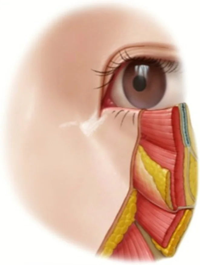
From the processes to the indication: selecting the candidate
Once the four processes are identified, the next question is which predominates, since the facelift resolves only one. The procedure repositions tissue: it corrects effectively what has been displaced and alters neither what is absent nor what has changed in quality. The indication therefore depends less on age or degree of laxity than on the nature of the defect. The table sets out the four profiles most frequently seen in consultation, with the examination finding that identifies each and the reason the therapeutic answer differs. The last three are not rejected patients but patients whose treatment sequence begins elsewhere.
Clinical profiles and therapeutic response
| Profile | Examination finding | Rationale |
|---|---|---|
| Direct indication | Competent skeletal structure — projected chin and malar eminence — with moderate nasolabial and nasojugal sulci and skin without advanced photoageing | The defect is exclusively positional, which is what repositioning corrects |
| Chin retrusion | Obtuse cervicomental angle with an accentuated melolabial sulcus | The deficit is of skeletal support: genioplasty or orthofacial surgery should be considered before the facelift or in the same procedure |
| Malar hypoplasia | Accentuated nasolabial sulcus over a flat midface | The deficit is volumetric: malar filling with hyaluronic acid or calcium hydroxyapatite, or fat grafting |
| Advanced cutaneous deterioration | Intense photoageing with deep static wrinkles | The facelift repositions but does not alter dermal quality: it requires associated or prior resurfacing, by peel or laser |
The operative definition of the candidate
Selection can be reduced to a single criterion: a patient with sufficient skeletal support whose medialisation of the cutaneous-adipose tissue arises from ligamentous laxity and not from absent underlying bone. The distinction is decisive. Where tissue has medialised for want of skeletal support, traction reproduces the same displacement within months, since the cause is unaltered. Where it has medialised because the ligaments have lost competence, repositioning acts on the mechanism and the result holds. The consultation should end with that question answered.
The midface: four variables that order the decision
The midface concentrates the greatest difficulty in facial rejuvenation, and the reason is not a scarcity of techniques but their abundance: transtemporal lift, transblepharoplasty approach, rhytidectomy with malar repositioning, fat grafting, implants and fillers all address the same region, so that without a classification the choice lacks foundation. Jacono and Rousso proposed a system assessing four variables by examination and preoperative photography: volume loss, midfacial descent, loss of elasticity and the presence of a negative orbital vector or hypoplastic malar eminence. Its value lies in each variable pointing towards a different kind of manoeuvre, their combination defining the plan.
Determining the orbital vector
It is the most objectifiable of the four variables, and the reference is the one described by Jelks and Jelks. With the patient in natural head position, a vertical is drawn through the corneal apex — the most anterior point of the globe — and compared with the position of the infraorbital rim. If the rim lies anterior to that vertical the orbital vector is positive; if they coincide, neutral; and if it lies posterior, negative. A negative vector expresses a deficit of skeletal projection, whence its two consequences: traction does not correct it, so volume or support must be added, and it predisposes to lower eyelid malposition after blepharoplasty.
Midface: reposition, add volume or both
Does the infraorbital rim lie behind the vertical drawn through the corneal apex?
Does tissue descent or compartment atrophy predominate?
Facial analysis: thirds and proportions
Assessment of the midface rests on analysis of the whole: symmetry, the proportion between height and width, and profile. In frontal view the face divides into thirds: the upper, from the hairline to the lower border of the brows; the middle, to the nasal base; and the lower, to the chin border. In a balanced proportion the three are equivalent. Within the lower third a second proportion applies: one third from the nasal base to the oral commissure and two thirds from the commissure to the chin. The scope of these canons deserves precision: they are not historical constants, they have varied across periods and the current reference tends towards a biprotrusive profile, so they serve to identify deviations rather than to impose a model. The full development, with its lines and angles, is found in Facial analysis: lines, angles and proportions.
Cervical assessment
The lower third and the neck are assessed by the same criteria of proportion, but they also have an explicitly described objective. Ellenbogen and Karlin defined the five visual criteria characterising a youthful neck, and their value is twofold: they set the surgical objective and provide concrete language in which to explain it to the patient. The anatomy underlying each of them — the cervical layers, the two fat compartments and the submuscular structures — is developed in Cervical anatomy for the facial surgeon.
With the objective defined, examination must attribute each deviation to the responsible structure, because each is corrected by a different manoeuvre and none substitutes for another. The manoeuvres below are performed in this order, from superficial to deep.
Manoeuvres of the cervical examination
- Pinch with swallowingPinch the submental tissue and ask the patient to swallow. If what is held corresponds to skin with subcutaneous tissue, the fat is supraplatysmal; if the tissue escapes during swallowing, it corresponds to lax platysma or subplatysmal fat
- Measuring the pinched foldOnce the tissue is confirmed supraplatysmal, the thickness of the pinched fold is measured between the fingers. Above 3 cm the volume suffices for liposuction to alter the contour. Below 3 cm the superficial layer is thin and the volume arises from the deep plane: aspirating it does not correct the contour and leaves the skin adherent to the platysma, with retraction and irregularity
- Platysmal bandsDistinguish between dynamic bands, appearing only on platysmal contraction, and adynamic bands, present at rest. Dynamic bands express muscular hyperfunction and are treated by partially dividing the band — transection; adynamic bands express loss of midline support and are treated by approximating both bellies with a corset suture, which restores continuity without excising muscle
- Submandibular glandsPalpate for glandular ptosis, which conditions the repositioning vector and the design of the platysmaplasty. The technical development is in Platysmaplasty and cervical contour
- Position of the hyoidA low, anterior hyoid limits the achievable cervicomental angle irrespective of the technique used. It is the only finding of the cervical examination that admits no correction, and must therefore be recorded in the preoperative information
What examination does not show: the directed history
Examination localises the anatomical defect but says nothing of healing capacity or bleeding risk, which condition the result as much as technique does. That information comes from the history, and five items are worth asking about explicitly because the patient does not usually mention them unaided.
The five items of history and their bearing
Each compromises the result by a different mechanism. Smoking is the factor that most increases the risk of flap necrosis. Haemostatic disorders and antiplatelet or anticoagulant therapy lead to haematoma, the most frequent complication of this surgery. Diabetes mellitus affects healing and the risk of infection. And a tendency to hypertrophic or keloid scarring is decisive in a procedure whose result depends on scars remaining concealed; its management is developed in Facial scar revision. Identifying them in consultation allows the patient to be optimised, the operation deferred or the indication withdrawn.
Closing the consultation: information and record
With examination and history gathered, the indication must be formulated and documented. Dissatisfaction after aesthetic surgery correlates with the distance between the expected and the obtained result more than with the technical quality of the operation, so preoperative information forms part of the procedure rather than its preamble. It must cover three headings: the foreseeable result, its limits and the possible complications. The aim is not to persuade but to establish a shared understanding of what is attainable. One question orders the conversation effectively: whether the patient seeks a modification of their features or the restitution of their own appearance at an earlier point, since these correspond to two distinct surgical propositions. What is agreed is then documented through the photographic record and written note of what will not change.
Minimum content of the preoperative record
- Preexisting asymmetries, demonstrated in the mirror and documented. They constitute the object of most late complaints.
- The elements that will not change: dermal quality, deep static wrinkles and the cervicomental angle when the hyoid is low.
- The complementary procedures proposed and their rationale — fat grafting, genioplasty, resurfacing — with express record if the patient declines them.
- The standardised photographic record, which also serves as objective reference in case of later dispute.
The conclusion that orders the indication
The whole analysis converges on one operative question: what proportion of the problem corresponds to descent and what proportion to volumetric atrophy. Where descent predominates, the manoeuvre is to reposition; where atrophy predominates, to restore volume; and in most patients beyond fifty, both within the same operation. On this depends whether a technically correct facelift proves unsatisfactory: with ptosis corrected and atrophy untreated, the volumetric component of ageing persists. The technical translation of that decision is developed in Planes and vectors in facelift surgery.
References
- 1.Vaquero Martínez P. Cirugía de lifting. En: Manual de la Sociedad Española de Cirugía Oral y Maxilofacial y de Cabeza y Cuello. SECOM CyC; 2025.
- 2.Jelks GW, Jelks EB. Preoperative evaluation of the blepharoplasty patient: bypassing the pitfalls. Clin Plast Surg. 1993;20(2):213-223.
- 3.Yaremchuk MJ. Infraorbital rim augmentation. Plast Reconstr Surg. 2001;107(6):1585-1592.
- 4.Moorrees CFA, Kean MR. Natural head position, a basic consideration in the interpretation of cephalometric radiographs. Am J Phys Anthropol. 1958;16(2):213-234.
- 5.Jacono AA, Rousso JJ. An algorithmic approach to multimodality midfacial rejuvenation using a new classification system for midfacial aging. Clin Plast Surg. 2015;42(1):17-32.
- 6.Rohrich RJ, Pessa JE. The fat compartments of the face: anatomy and clinical implications for cosmetic surgery. Plast Reconstr Surg. 2007;119(7):2219-2227.
- 7.Mendelson B, Wong CH. Changes in the facial skeleton with aging: implications and clinical applications in facial rejuvenation. Aesthetic Plast Surg. 2012;36(4):753-760.
- 8.Ellenbogen R, Karlin JV. Visual criteria for success in restoring the youthful neck. Plast Reconstr Surg. 1980;66(6):826-837.
Related specialty: Facial Lifting & Rejuvenation