Platysmaplasty and cervical contour — Management of the subplatysmal plane
The restricted indication for cervical liposuction, management of the subplatysmal plane, medial and lateral platysmaplasty step by step, and the three manoeuvres that open the cervicomental angle.
Content intended for healthcare professionals. It does not replace clinical assessment and is not written as patient information.
Key points
- In the non-obese patient, most of the cervical volume lies beneath the platysma, so the superficial plane rarely contains the excess that is to be addressed.
- Cervical subcutaneous tissue contributes to a youthful appearance: emptying it does not slim the neck but produces retraction and transparency of the underlying muscle.
- Three manoeuvres open the cervicomental angle and are performed systematically: myotomy of the anterior digastric belly, prehyoid fasciotomy and partial transection of the bands.
- Excision must be limited to interplatysmal fat and not reach the interdigastric compartment: removing the latter fuses skin to the digastric bellies and produces a deformity difficult to correct.
- The physiological cervical contour presents greater central than lateral volume; pursuing a uniform concavity produces a result recognisable as surgical.
The change of plane that defines current technique
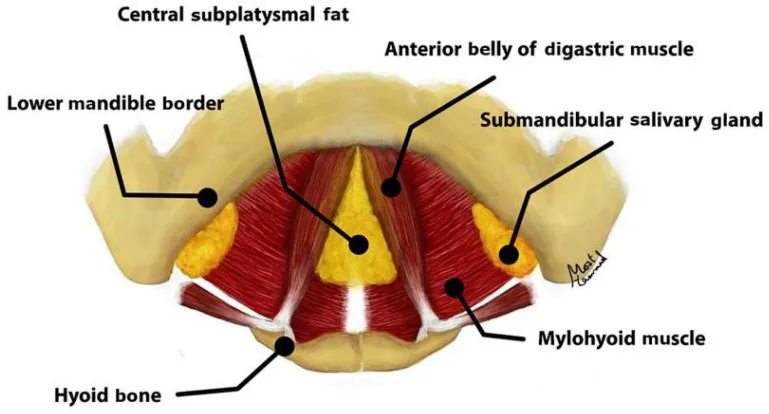
Cervical surgery developed for decades on the supraplatysmal plane, through liposuction and skin excision. The description of the two fat compartments and of the submuscular structures — developed in the note on cervical anatomy — shifted the focus to the deep plane, and that shift is what separates a natural result from an emptied one. The preoperative check is direct: pinch the submental tissue and ask the patient to swallow. If the tissue held between the fingers corresponds to skin with subcutaneous tissue, supraplatysmal fat amenable to treatment is present; if it escapes during swallowing, the excess is deep or corresponds to muscular laxity, and liposuction does not reach it.
Manoeuvres of restricted and of systematic indication
From the anatomy described follows a hierarchy of indications that differs from classical practice. Three manoeuvres formerly routine now have a restricted indication, and three that were omitted are performed systematically. The table below sets out each with its criterion and rationale.
Criterion of indication by manoeuvre
| Manoeuvre | Indication | Rationale |
|---|---|---|
| Cervical liposuction | Restricted | The predominant volume is subplatysmal. Emptying the superficial plane accentuates the bands and produces cutaneous retraction. Indicated when the pinched fold exceeds 3 cm and corresponds to skin with subcutaneous tissue |
| Platysmectomy | Restricted | Dynamic bands are corrected by muscular transection and adynamic bands by medial corset suture. Muscular excision adds tension without adding correction. Indicated only for markedly lax bands |
| Extensive central lipectomy | Contraindicated | Excision of interdigastric fat fuses the skin to the muscle bellies and produces the cobra deformity |
| Digastric myotomy | Systematic | The anterior belly is frequently hypertrophic or lax and generates submental fullness. The redundant portion is excised |
| Prehyoid fasciotomy | Systematic | It releases the medial platysma from the hyoid and infrahyoid musculature, which permits greater tension in the plication and contributes to closing the angle |
| Partial transection of the bands | Systematic | Applicable both to the central band generated by the platysmaplasty itself and to dynamic bands from hyperfunction. Always partial: complete division produces a palpable discontinuity |
Medial platysmaplasty
- IncisionSubmental, some 2 cm, placed 2 mm caudal to the submental crease, where the scar is less apparent, with a cold blade
- Supraplatysmal planeDissect to the sternocleidomastoid and the level of the thyroid cartilage, running along the entire mandibular border and releasing the mandibular ligament with scissors
- Opening the platysmaIn the midline, where the muscle is usually absent. Beneath appear the anterior digastric bellies, which delimit the deep plane
- Subplatysmal planeDissect with scissors held vertically until the submandibular glands are identified, releasing the mandibular ligament from this access as well
- LipectomyInterplatysmal excision as far as the plane of the digastrics, preserving the interdigastric fat intact
- Digastric and fasciaMyotomy of the anterior belly with bipolar excision of the redundant portion, and transverse prehyoid fasciotomy of the deep cervical fascia — not of the musculature — after palpating the hyoid
- Submandibular glandIf indicated, reduction of the superficial glandular lobe, preserving the facial vessels. The vein lies anterior to the artery, which constitutes the reference on approach
- PlicationSuture of the platysma with 4/0 absorbable material in cross stitches, submental and cervical. A running suture is not advised since it generates a visible central band
Indication and extent of transection
There are two transections with distinct indications. That of the central prehyoid band is performed when, with the neck in hyperextension, the band presents an obtuse rather than acute profile; it is the band generated by the platysmaplasty itself. That of the dynamic band at the level of the thyroid cartilage is performed when preoperative examination identified hyperdynamic bands. In both cases the same criterion applies: partial transection and never complete division, with diathermy centrally and with scissors against the muscle laterally, over approximately one centimetre and directed laterally and superiorly. Complete division does not improve the angle and produces a palpable discontinuity.
Lateral platysmaplasty
- Supraplatysmal dissectionFrom the S-shaped incision, to the sternocleidomastoid and the level of the thyroid cartilage, running along the entire mandibular border and releasing the mandibular ligament
- Point of entryThe gonion for the cervical ligament, and over the belly of the sternocleidomastoid to preserve the external jugular vein and the great auricular nerve. Infiltrate the subplatysmal plane beforehand
- Subplatysmal planeDissect to the facial artery and release the platysma from the sternocleidomastoid as far as the thyroid cartilage. The cervical ligament presents the greatest technical difficulty of this sequence
- Parotid tailIncision at the anterior border of the infralobular sternocleidomastoid, dissection of the supraparotid SMAS and excision of the tail. Preserve continuity between platysma and facial SMAS, whose division removes tension over the gonion and with it definition
- SuspensionTo the mastoid periosteum with 2/0 absorbable material, at 80 degrees, with a mattress stitch on the platysma to distribute tension. Two points of fixation
- ClosureIrrigation, placement of drains and tension-free skin closure, with passive excision of the excess
The thickness of the cervical flap
The principle established for the facial flap applies to the cervical flap with greater reason: a layer of adipose tissue must be preserved adherent to the skin flap. Dissection flush with the dermis produces two findings appreciable in the medium term: darkening of the skin through transparency of the underlying platysma and contour irregularity. The consequence runs contrary to the objective pursued, and is worth stating explicitly: reduction of cervical volume is obtained in the subplatysmal plane, not by thinning the flap.
Technical precautions
- During glandular reduction, the facial vein lies anterior to the artery and constitutes the reference on approaching the plane.
- Placing the platysmal opening over the belly of the sternocleidomastoid simultaneously preserves the external jugular vein and the great auricular nerve.
- The external jugular vein remains adherent to the deep plane during platysmal dissection and is not elevated with the flap.
- In a technique limited to the neck, it is advisable to complete the contralateral side before closure so that both may be compared and balanced.
- Quilting stitches in the submandibular region at the level of the gonion: they obliterate dead space and reduce the risk of haematoma and seroma. They are removed on the third day.
The objective and its limit
The criteria of Ellenbogen and Karlin define the achievable objective and are worth keeping in mind during the operation: a defined mandibular border, subhyoid depression, relief of the thyroid cartilage, visible anterior border of the sternocleidomastoid and a cervicomental angle between 105 and 120 degrees. To these is added the anatomical limitation that must be explained before operating: a low, anterior hyoid sets an upper limit to the achievable angle, irrespective of the technique applied. Examining it in consultation moves that limitation to the point where it can be integrated into the patient’s expectation.
References
- 1.Vaquero Martínez P. Cirugía de lifting. En: Manual de la Sociedad Española de Cirugía Oral y Maxilofacial y de Cabeza y Cuello. SECOM CyC; 2025.
- 2.Feldman JJ. Corset platysmaplasty. Plast Reconstr Surg. 1990;85(3):333-343.
- 3.Ellenbogen R, Karlin JV. Visual criteria for success in restoring the youthful neck. Plast Reconstr Surg. 1980;66(6):826-837.
- 4.Baker DC. Face lift with submandibular gland and digastric muscle resection: radical neck rhytidectomy. Aesthet Surg J. 2006;26(1):85-92.
- 5.Guyuron B, Jackowe D, Iamphongsai S. Basket submandibular gland suspension. Plast Reconstr Surg. 2008;122(3):938-943.
- 6.Mendelson BC, Tutino R. Submandibular gland reduction in aesthetic surgery of the neck. Plast Reconstr Surg. 2015;136(3):463-471.
- 7.Bray D. Extended composite approach to deep plane face lifting with deep contouring of the neck. Facial Plast Surg. 2024;40(6):750-765.
- 8.Boyd CJ, Ceradini DJ. Current trends in facelift and necklift procedures. J Clin Med. 2025;14(12):4273.
- 9.Talei B. The detailed anatomy of the deep plane of the face and neck with current nomenclature. Facial Plast Surg. 2024;40(6):680-686.
Related specialty: Facial Lifting & Rejuvenation