Upper blepharoplasty — Marking, technique and variants
What defines a youthful upper lid, how to rule out ptosis and brow descent before marking, millimetre-by-millimetre marking and the full surgical sequence with its variants.
Content intended for healthcare professionals. It does not replace clinical assessment and is not written as patient information.
Key points
- The goal is not a lid without skin: it is to reproduce the youthful lid — little show, lateral and central convexity, slight medial concavity.
- Resect skin and, where indicated, the medial pad. Muscle and central fat are not resected routinely: that is the route to the hollowed eye.
- Before marking, rule out two things mistaken for skin excess: eyelid ptosis and brow descent.
- The non-negotiable measurement: leave 10 mm of skin between the upper incision and the orbital rim. Below that, lagophthalmos appears.
- The only justified muscle resection is in the patient with ptosis and a hypertrophic orbicularis, and always without crossing the septum.
Anatomy I — What is being reproduced
It is worth having the goal in mind before picking up the marker, because upper blepharoplasty is surgery of shape, not of quantity. The youthful lid has four constant features. First, little lid show between crease and lashes, more marked in women than in men. Second, convexity in the lateral and central thirds: the youthful lid is full, not empty. Third, a slight medial concavity, the only area where some hollow is acceptable. And fourth, an elevated brow tail, peaking between the lateral limbus and lateral canthus in women, while in men the brow is flatter and lower. The whole indication follows from these four: resect what hides the convexity — skin — and preserve what creates it — muscle and central fat.
Anatomy II — The layers and what lies behind the septum
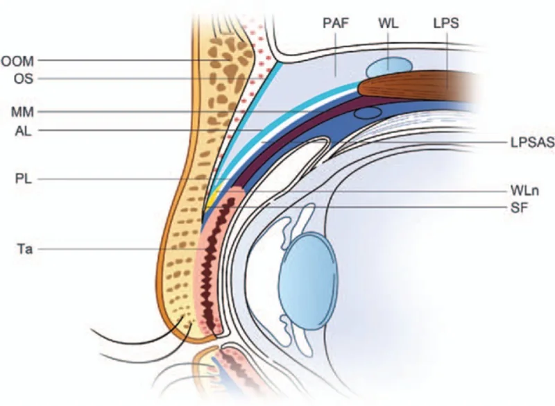
From surface to depth: skin, orbicularis, orbital septum, preaponeurotic fat, levator aponeurosis, Müller’s muscle, tarsus and conjunctiva. The subcutaneous plane is extremely thin — eyelid skin is the thinnest in the body — which demands precise dissection and explains why the marking is made by stippling rather than a continuous line. Behind the septum lie two fat compartments, not three as in the lower lid: the medial (nasal), characteristically paler, whitish and more fibrous, and the central or preaponeurotic, more yellow owing to its higher carotenoid content. That colour difference is the practical landmark distinguishing them in the field. And one fact governs the safety of this operation: the levator aponeurosis lies immediately behind the preaponeurotic fat. Hence the rule: do not cross the septum unless deliberately going for fat, and do so through a small, controlled opening.
Examination: three diagnoses that get confused
The patient says “my eyelid feels heavy”, but that can come from three different places and only one is solved by resecting skin. Dermatochalasis is the blepharoplasty indication. Eyelid ptosis is different: the lid margin covers part of the pupil because the levator has lost power or its aponeurosis has disinserted, objectified by MRD1 and by levator function. And brow descent mimics skin excess because it drags forehead tissue downward; it is uncovered by lifting the brow with a finger to its correct position and seeing how much skin is truly redundant then. Operating on the lid when it is the brow that has dropped does not fix the problem and may worsen it, because closing the incision pulls the brow tail further down.
Lid, brow or ptosis?
Is MRD1 under 4 mm, or does the lid cover part of the pupil?
Lifting the brow with a finger to its correct position, does most of the skin excess disappear?
The marking, millimetre by millimetre
The marking is done with the patient sitting and awake, before infiltration, and by stippling: the skin is so thin that a continuous line distorts when stretched. Excess skin is grasped with bayonet or Green forceps. Lower line: stretch the skin upward and draw a line parallel to the lid margin along the lid crease, at 8 mm in men and 10 mm in women — a wider upper lid reads as more feminine. Upper line: now stretch the redundant skin downward and draw a second parallel line above it, always leaving 10 mm of skin up to the orbital rim. The one-centimetre rule still holds as a reference, adapted to each anatomy. The two ends have their own shape: medially the lines meet in a downward-pointing apex, and here one must be especially sparing, because removing much tissue at the inner angle causes retraction and webbing; laterally they meet in an upward apex following a static crease of the periorbital rhytidosis.
The two measurements that are not negotiable
10 mm of skin between the upper incision and the orbital rim: below that reserve the lid does not close and lagophthalmos appears, whose correction requires a retroauricular skin graft. And restraint at the medial end: the inner angle tolerates resection poorly and responds with retraction and webbing. In both cases the error is made at the marking stage, not in theatre, which is why the drawing is checked with the patient sitting and eyes open before any infiltration.
Surgical sequence
- 1. PositionSurgeon at the head of the patient, with symmetric view of both lids
- 2. AnaesthesiaEntry point with a fine needle and a blunt 27 G, 38 mm cannula in the subcutaneous plane, visible under the skin, with articaine and adrenaline
- 3. IncisionFine-tip electrocautery along the marking from lateral to medial; assistant with two gauzes providing counter-traction
- 4. Skin resectionDissection in the very thin subcutaneous plane, coagulating any bleeding vessel in the deep plane
- 5. Muscle (optional)Only if indicated: a strip of orbicularis. Never cross the septum at this step
- 6. FatSmall septal incision over the compartment to be treated; assistant applying globe pressure to deliver the pad
- 7. HaemostasisRetract the pad, clamp with a mosquito, trim with scissors and coagulate the stump above the clamp before releasing
- 8. ClosureMuscle layer with 5/0 Vicryl covering the septal opening — the septum is not sutured —; skin with a 5/0 prolene subcuticular suture, medial to lateral
Why muscle and central fat are not routinely resected
Both create the convexity that defines a youthful lid. Removing them produces the hollowed eye: a skeletonised lid with a visible orbital rim, which ages the gaze far more than the redundant skin ever did. It is a complication that is hard to correct and requires fat grafting with a less predictable result than the original operation. Fat resection is therefore limited to the medial pad when it protrudes, is partial, and never empties the compartment. Muscle resection is reserved for the patient combining ptosis with a hypertrophic orbicularis, and consists of a strip, not a whole layer.
Variants according to the finding
| Finding | What is added | Caveat |
|---|---|---|
| Xanthelasma | Extended resection of the skin ellipse, or electrocautery | The only treatment, but with a substantial recurrence risk: say so beforehand |
| Hypertrophic orbicularis with ptosis | Resection of a strip of orbicularis | The only indication for muscle resection. Without ptosis, the muscle is left alone |
| Risk of brow tail descent (typical in women) | Lateral suspension stitch: divide the most lateral orbicularis and anchor tarsal portion, orbital rim periosteum and ciliary portion with two 5/0 Vicryl stitches | Reduces the likelihood of later needing a brow lift |
| Brow tail already dropped | Botulinum toxin to the lateral orbicularis, or a brow tail lift | If the component is marked, resecting skin without addressing it worsens the result |
| Hollowed eye | Autologous fat grafting: 0.9 mm Tulip cannula, 0.5-1 cc, supraperiosteal plane at the superior orbital rim | Never in the lid itself, always at the rim. Massage afterwards to avoid irregularities |
The lateral dog-ear and how to solve it
At the end of closure a redundant fold may remain at the lateral end — the dog-ear. It does not flatten on its own, so it is dealt with during the same procedure: extend the scar with Converse, Iris or Stevens scissors along the same periorbital crease used to end the incision. That lateral extension benefits from safety stitches of fast chromic catgut or 6/0 Vicryl rapide, since it is the area under most tension. Anticipating it at the marking stage — extending the lateral end from the outset when the excess is obvious — saves this step.
Postoperative regimen
- On finishingWhile the second eye is operated, the first is covered with cold moist gauze. Iodine applied with a fine tip and adhesive strips
- ExpectationWarn of pain and deformity for two weeks: the least well tolerated phase and the one generating most calls
- AnalgesiaParacetamol 1 g + dexketoprofen 25 mg + prednisone 30 mg for one week
- Eye careAntibiotic ointment every 12 h for one week, alternating with artificial tears every 8 h
- SuturesRemoved at 7 days
The two complications to know how to solve
Retroseptal haematoma: intense progressive eye pain, proptosis or visual loss mandate lateral canthotomy with cantholysis without delay, without waiting for imaging. This is why the fat pedicle is always coagulated before release. Lagophthalmos from excessive skin resection: the lid does not close, the cornea is exposed, and the solution is not to wait but to replace skin with a retroauricular graft. Both complications are prevented at specific steps — haemostasis at step 7, the 10 mm reserve at marking — not by general surgical skill.
References
- 1.Demetriades NC, Madnani DD. Periorbital Rejuvenation with Application of Fat Transfer. Atlas Oral Maxillofac Surg Clin North Am. 2018;26(1):69-75.
- 2.Alghoul M, Codner MA. Retaining Ligaments of the Face. Aesthet Surg J. 2013;33(6):769-782.
- 3.American Academy of Ophthalmology. Basic and Clinical Science Course, Section 7: Oculofacial Plastic and Orbital Surgery. San Francisco: AAO; 2023.
- 4.Gurtner GC, Neligan PC, editors. Plastic Surgery. Volume 2: Aesthetic Surgery. 4th ed. Edinburgh: Elsevier; 2018.
Related specialty: Blepharoplasty