Cutaneous wound healing — From the classic phases to reverse-engineering the scar
The phases and their mediators, the factors that derail healing, and why the fetus does not scar: the Engrailed-1 lineage, mechanotransduction, and what can already be blocked.
Content intended for healthcare professionals. It does not replace clinical assessment and is not written as patient information.
Key points
- The scar is not the inevitable price of repair: it is the output of a specific cellular programme that has been identified and is beginning to be blockable.
- The pilosebaceous unit is the skin’s stem cell reservoir. This is why hair-bearing skin recovers from resurfacing far better than non-hair-bearing skin.
- Mechanical tension is the switch that activates the fibrotic lineage. Hence all the classic rules — close without tension, direct it horizontally, obliterate dead space — now have a molecular explanation.
- The fetus does not scar because its fibroblasts belong to another lineage and its myofibroblasts are only transient. Adult oral mucosa behaves similarly.
- Moist occlusion accelerates epithelialisation compared with a dry scab. It dates from the 1960s and remains among the most cost-effective things one can do for a wound.
Why skin anatomy decides the result
The epidermis provides the barrier and the colour, and contains no collagen: hence a freshly epithelialised wound has minimal mechanical strength even though it looks closed. Its growth capacity is enormous and seals a fresh wound within hours. If the injury spares the basal layer — an abrasion, a superficial burn — healing occurs without scarring, though it may leave dyschromia because the melanocytes live there. The dermis provides thickness and toughness, and its collagen takes far longer to lay down than the epithelium takes to close. That asymmetry between fast closure and slow strength explains most early dehiscences.
The pilosebaceous unit explains why some areas recover and others do not
Follicles and sebaceous glands are not mere appendages: they are the progenitor reservoir from which skin regenerates after injury, with their own vascularity and innervation. Specific markers of those progenitors have been identified — Lgr6 among them — and their presence explains an everyday clinical observation: hair-bearing skin tolerates and recovers from resurfacing far better than non-hair-bearing skin. It is why glabrous areas demand more conservative parameters, and why a graft from a hairy area behaves differently from one taken from a hairless one.
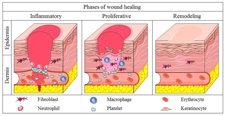
The phases and what happens in each
| Phase | What happens | When |
|---|---|---|
| Vasoconstriction | Immediate response to injury, before anything else | 5-10 minutes |
| Inflammatory | Coagulation cascade, platelet aggregation and release of PDGF, VEGF, EGF, IGF-1, TGF-β and FGF-2. Then vasodilation from histamine and serotonin | Peak permeability in the first 48-72 h |
| Proliferative | Angiogenesis driven by macrophages, platelets, lymphocytes and mast cells. Low oxygen tension is what triggers angiogenic factors | From day 4 |
| Remodelling | Replacement of collagen III by I, fibre realignment and progressive gain in strength | Months. The scar is not finished at 6 weeks |
Hypoxia is a signal, not only a problem
One physiological detail has practical consequences: low oxygen tension in the bed is what prompts macrophages to release angiogenic factors. When oxygenated blood arrives, that stimulus falls and neovascularisation slows. It is a self-regulating system. The clinical reading is not that ischaemia is desirable — an ischaemic wound does not heal — but that the oxygen gradient is information, and an artificially and continuously hyperoxygenated wound does not necessarily heal better.
Factors that derail healing
- Local: ischaemia, infection, tissue trauma, retained foreign body, desiccation, venous stasis, lymphoedema, radiation and previous surgery in the area.
- Metabolic and endocrine: diabetes, chronic renal failure, Cushing syndrome, hypothyroidism and metabolic syndrome.
- Cardiovascular: atherosclerosis, heart failure, hypertension, vasculitis and microvasculopathy.
- Drugs and substances: tobacco, glucocorticoids, antineoplastics, colchicine, penicillamine, anticoagulants, antiangiogenics, and at high dose salicylates, NSAIDs, vitamin E, vitamin A and zinc sulfate.
- Systemic: malnutrition, immunodeficiency, liver disease, chronic lung disease, malignancy and hereditary collagen disorders such as Ehlers-Danlos or Marfan.
A note on high doses
That list rewards careful reading: vitamin A, vitamin E and zinc appear among the factors that impair healing, while also being used as supplements to improve it. This is not contradictory: the issue is dose. In deficiency, supplementing helps; at supraphysiological doses, vitamin E interferes with platelet aggregation and collagen synthesis, and high-dose zinc competes with copper absorption, which collagen cross-linking requires. Vitamin A is the special case: it counteracts the inhibitory effect of corticosteroids, which makes it useful in the steroid-treated patient and dispensable in everyone else.
Reverse engineering: why the fetus leaves no scar
The starting observation is old: fetal wounds heal without scarring. The differences from the adult are described — extracellular matrix composition, closure dynamics, gene expression — but two are especially revealing. First: fetal myofibroblasts are only transient, which makes those wounds far less susceptible to cicatricial contracture. Second is an experiment that points straight at the mechanism: adding interleukin-6 to a fetal wound produces a scar where normally there would be none. That is, scarlessness is not a magical property of fetal tissue but the result of a different inflammatory environment that can be altered. One adult model behaves similarly and we see it daily: oral mucosa heals fast and with minimal scarring, with fewer immune and profibrotic mediators.
The Engrailed-1 lineage: the scar has a responsible cell
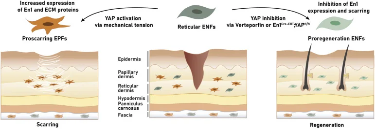
The conceptual leap of the past decade is that not all fibroblasts are alike. Two populations have been identified by their history of expressing the Engrailed-1 gene: those that have expressed it at some point, profibrotic, and those that never have. The former produce the scar. And the decisive finding is that the gene can be activated after birth: following a wound, fibroblasts that had never expressed it switch it on, convert to the profibrotic phenotype, dominate the bed and deposit excess collagen I. The scar, therefore, is not a destiny: it is a cellular conversion occurring during repair.
And the switch is mechanical
What activates Engrailed-1 is mechanical tension, through canonical mechanotransduction and the YAP factor. Blocking that pathway with verteporfin in animal models produces something that until recently sounded like science fiction: regeneration instead of scarring, with recovery of hair follicles, glands, matrix ultrastructure and normal mechanical strength. Strategies for targeted silencing of the gene with a single topical administration have also been published. For the surgeon there is an immediate conclusion, applicable today without waiting for any drug: the classic rules of closing without tension, orienting the incision and offloading the wound are not surgical aesthetics, they are direct intervention on the molecular switch of the scar.
What can be done today in every wound
- DesignOrient the incision along tension lines and direct the resulting tension away from mobile structures
- TechniqueHalsted’s principles still hold: gentle tissue handling, anatomical dissection, careful haemostasis, obliteration of dead space and no tension
- ClosureThe load is borne by the deep layer; skin sutures only appose. Remove sutures early on the face to avoid track marks
- EnvironmentMoist occlusion: it accelerates epithelialisation compared with a dry scab, where cell migration and survival are impaired
- OffloadingAdhesive strips, silicone dressings and pressure maintained for months, not weeks: remodelling remains active long after discharge
- Patient factorsSmoking cessation, glycaemic control and review of drugs and supplements before surgery
References
- 1.Frodel JL, Brenner MJ. Wound healing. In: Papel ID, ed. Facial Plastic and Reconstructive Surgery. 4th ed. New York: Thieme; 2016.
- 2.Mascharak S, desJardins-Park HE, Davitt MF, et al. Preventing Engrailed-1 activation in fibroblasts yields wound regeneration without scarring. Science. 2021;372(6540):eaba2374.
- 3.Winter GD, Scales JT. Effect of air drying and dressings on the surface of a wound. Nature. 1963;197:91-92.
- 4.Snyder RJ, Lantis J, Kirsner RS, et al. Macrophages: a review of their role in wound healing and their therapeutic use. Wound Repair Regen. 2016;24(4):613-629.
Related specialty: Complex Facial Reconstruction