Brow lift — Why the tail falls, where the nerve runs and which approach to choose
The anatomy explaining why the brow falls laterally, the frontotemporal branch layer by layer, the three resuspension vectors and the three approaches — temporal, frontotemporal and direct — with their indications.
Content intended for healthcare professionals. It does not replace clinical assessment and is not written as patient information.
Key points
- The only brow elevator is frontalis, and it ends at the temporal crest. Lateral to that there is only a depressor, orbicularis. That asymmetry explains why the brow falls sooner and further at its tail.
- A dropped brow mimics upper eyelid ptosis that blepharoplasty does not correct: it masks it and sometimes worsens it. Telling them apart before operating is the step that saves most results.
- The brow is not resuspended along one vector but three: medial oblique, central following the temporal crest and lateral almost vertical. A pure vertical vector across the whole brow is what produces the startled look.
- Facial nerve safety is a matter of plane, not dexterity: the frontotemporal branch always travels in the superficial flap, and there are two planes where it cannot be.
- What can be promised has been measured: the most recent meta-analysis gives 3.8 mm of lateral elevation, 3.0 mm central and 2.4 mm medial at nearly two years. It is a millimetre operation, not a centimetre one.
Brow ageing is read in the tail
The brow does not fall uniformly: it falls laterally, and that is the diagnosis. Three reasons add up. The first is muscular: frontalis, the only elevator, does not reach beyond the temporal crest, so the lateral brow has a depressor but no elevator. The second is ligamentous: the superficial temporal fascia is firmly bound to skin but loosely bound to the deep temporal fascia, except at the adhesion zone; that sector slides over the deep plane with little holding it. The third is skeletal: resorption of the orbital rim withdraws the support the brow rested on. The result has two clinical consequences: a sad expression and a pseudoptosis of the upper eyelid.
Who raises the brow and who lowers it
| Muscle | Action | Innervation |
|---|---|---|
| Frontalis | The only elevator. It ends at the temporal crest: it does not elevate the brow tail | VII nerve, frontotemporal branch |
| Corrugators (2) | Depress and draw medially | VII nerve, frontotemporal branch |
| Depressor supercilii (2) | Depress the medial portion | VII nerve, frontotemporal branch |
| Procerus | Depresses glabella and interbrow | VII nerve, buccal branch |
| Orbicularis oculi | The only muscle lateral to the temporal crest, and it is a depressor | VII nerve |
Sensation of the area
- It depends on the trigeminal, V1 division: supraorbital and supratrochlear nerves.
- They are the reason the medial release is done with the finger and not with a sharp instrument.
- Injuring them paralyses nothing, but leaves scalp dysaesthesia, which the patient does notice and has not always been warned about.
The layers, depending on where we are
| Frontal region | Temporal region | |
|---|---|---|
| 1 | Skin and subcutaneous / scalp | Skin and subcutaneous / scalp |
| 2 | Superficial galea | Temporoparietal fascia (superficial temporal) → continuous with the SMAS |
| 3 | Frontalis muscle | Superficial layer of the deep temporal fascia → continuous with the masseteric-parotid fascia |
| 4 | Deep galea | Temporal fat, loose areolar tissue (intermediate fat pad) |
| 5 | Pericranium | Deep layer of the deep temporal fascia (both anchor to the zygomatic arch) |
| 6-7 | — | Temporalis muscle and deep fat pad; pericranium |
Where everything sticks: the adhesion zones
There are two points where the layers stop gliding and fuse, and they are exactly the ones to release if the resuspension is not to relapse. At the temporal crest and lateral orbital rim, the superficial temporal fascia continues into the superficial galea, and the deep galea and pericranium fuse with both leaves of the deep temporal fascia. At the supraorbital rim, pericranium and orbital septum fuse and continue into pericranium and galea. Translated into manoeuvres: the lateral orbital ligament and the lateral arcus marginalis are the brakes, and until they are released any traction merely tensions them and the result is lost as healing proceeds.
The frontotemporal branch, plane by plane
- 1. In the parotidIntraparotid
- 2. On exitingSub-SMAS plane, between the two temporal fasciae, alongside the temporal vessels
- 3. Over the zygomatic archDeep to the superficial temporal fascia, at the middle third of the arch
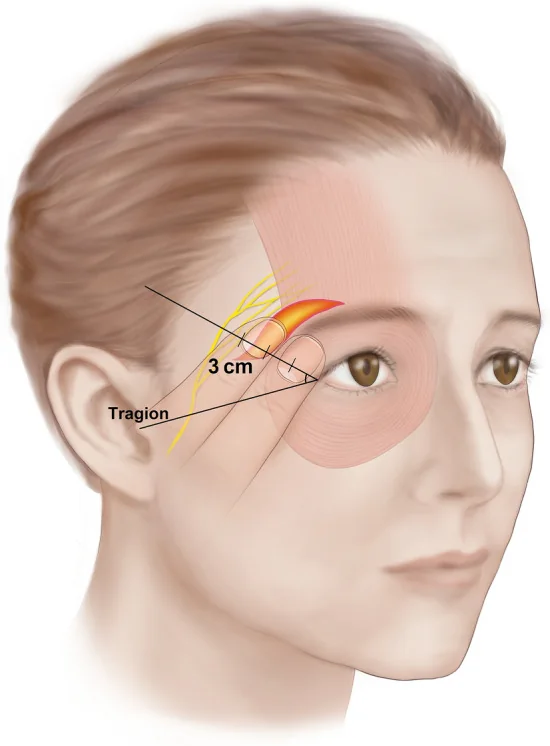
- 4. 1.5 cm above the archIt becomes superficial and travels within the superficial temporal fascia. Pitanguy’s line — earlobe to brow tail — marks its course
- 5. Once on the foreheadWithin the superficial galea, until it reaches frontalis on its deep surface
The two planes where the nerve cannot be
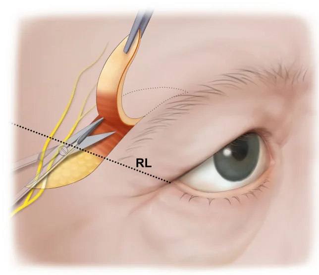
Two rules follow, and they solve the problem without needing to see the nerve. First: dissect deep to, or just superficial to, the deep temporal fascia — that unmistakable pearly white sheet — and the frontotemporal branch stays intact within the superficial flap, above the dissection. Second: dissect superficial to the superficial temporal fascia and the branch stays below. What is dangerous is exactly what lies between: the intra-superficial-fascial plane above the arch, which is where the nerve lives. Once the plane is chosen, the rest is patience.
The sentinel vein
Lateral to the lateral orbital ligament, along Pitanguy’s line, a perforating vein crosses from the deep to the superficial temporal fascia. It is not an obstacle: it is a warning. Its presence means the frontotemporal branch is immediately above, in the flap. Seeing it should change the tempo: from there on, blunt dissection, rotatory movements and no blind sharp manoeuvre. It is probably the most useful landmark of the whole temporal dissection.
What a normal brow is
- Man: a straight brow sitting on the supraorbital rim. Raising it feminises.
- Woman: an arched brow above the supraorbital rim, peaking towards the lateral canthus — classically at the lateral limbus.
- This is why in men caution with brow lift is advisable: as it descends with age, the male brow accentuates the supraorbital rim, which is a masculine feature, not a defect.
Before indicating: three questions in order
Is the pupil partly covered by the upper eyelid?
Phenylephrine test: does the aperture change after 2 minutes?
Is upper blepharoplasty enough or is a brow lift needed?
The commonest mistake
Correcting with upper blepharoplasty what is really a dropped brow. The excess eyelid skin is largely brow skin that has descended. Resecting it without repositioning the brow gives a cleaner eyelid and a brow that sits even lower, with less skin left to raise it later. Sequence matters: decide the brow first, then mark the eyelid — and if both are done in one procedure, mark the eyelid incision with the brow already elevated.
The three resuspension vectors
| Brow portion | Vector | Approximate angle |
|---|---|---|
| Medial | Oblique, laterally | 45° |
| Central | Following the temporal crest | 55° |
| Lateral (temporal) | Almost vertical | 80° |
Two goals, and one of them is not elevation
The goals of a brow lift are to reposition the brow over the orbital rim and to obtain a relaxed forehead. The second gets forgotten, and it is what separates a good result from a recognisable one: an overtightened brow, or a vertical vector applied to the whole brow instead of only its lateral portion, produces a permanently startled expression. Resuspension is not one upward pull, but three different pulls that respect the shape of the brow.
Temporal approach, step by step
- MarkingTemporal crest, Pitanguy’s line and incision line. The incision lies lateral to the crest if only the tail is to be repositioned, or on both sides if the whole brow is
- AnaesthesiaSedation and tumescent local anaesthesia. Part the hair with elastics and iodine gel or ointment, reapplied during surgery
- Incision2-3 cm inferolaterally from the temporal crest, cold blade, bevelled parallel to the follicles to avoid cicatricial alopecia. Through skin, subcutaneous tissue and superficial temporal fascia down to the deep temporal fascia
- Cranial dissectionRelease the whole frontotemporoparietal scalp: not a luxury but what prevents closure tension, and with it alopecia and dehiscence
- Medial dissectionInterfascial plane (leaving the deep temporal fascia below; it is avascular) up to the temporal crest adhesion zone, where one changes to the subperiosteal plane across the forehead
- Interbrow zoneRelease it from the contralateral approach, and perform the medial brow release with finger dissection, so as not to injure the supraorbital nerves or vessels
- Inferior dissectionRelease the lateral arcus marginalis to the mid-brow and the lateral orbital ligament: without this the resuspension relapses on healing
- How to dissectThe key: medial and caudal planes, slowly and with rotatory movements. Blunt dissector and non-cutting scissors; the instrument pushes and separates, it does not cut
How to fix the flap
| Method | How it is done | Note |
|---|---|---|
| Suture to deep temporal fascia | 3 sutures of long-lasting absorbable 2/0: mattress in the superficial temporal fascia and figure-of-eight in the deep | The default method |
| Bone fixation | With piezoelectric or bur, two cuts in the outer cortex parallel to the traction vector, leaving a cortical bridge between them through which the suture passes via the medullary bone. Bone wax if it bleeds | Rigid fixation with no permanent foreign material |
| Screws or tined devices | Titanium or absorbable, fixing the inferior flap | The patient may feel them. In the recent meta-analysis tined implants gave more lateral elevation but more dysaesthesia than suture |
Closure and adjunctive manoeuvres
- The traction vector follows the temporal crest. This rule governs fixation in both the temporal and frontotemporal approaches.
- Close the subcutaneous layer and then use staples, which cause less ischaemia and less cicatricial alopecia than a running suture; alternatively, a running locked 5/0 nylon.
- Transfixing sutures in the temporal area to prevent haematoma in the dissected space.
- Corrugator and procerus resection: medial incision in a forehead crease or in scalp, subperiosteal plane including the arcus marginalis while sparing the supraorbital and supratrochlear nerves, division with cautery and resuspension of the inferior flap to pericranium and deep galea.
- Blocking the medial depressors is an adjunct specific to the endoscopic technique; botulinum toxin serves the same purpose without surgery and also helps modulate the result.
Combined frontal and temporal approach
- MarkingTemporal crest, Pitanguy’s line, perpendicular frontal incision and parallel temporal incision
- Temporal routeAs in the temporal approach: interfascial plane to the crest, subperiosteal on the forehead, release of the lateral arcus marginalis and orbital ligament
- Frontal routeIncision down to bone and completion of the parietal and frontal dissection, posterior areas first and anterior after
- Medial sutureFrom both frontal incisions, a horizontal mattress of long-lasting absorbable 2/0 joining pericranium and lateral galea of one side to those of the other. It is tied while the assistant elevates the brows
- Lateral suturesFrom both temporal incisions, horizontal mattress sutures from inferior to superior flap, again tied with the brows elevated
- ClosureLow-pressure drain, running locked 5/0 nylon and temporal transfixing sutures
Direct brow lift: the technique dismissed too soon
Excising a spindle of skin just above the brow is the simplest and most precise manoeuvre: you see exactly how much is elevated and where. It is dismissed for fear of the scar, and that fear is founded in the medial region, less so laterally. It has very clear indications of its own: men, especially with a high forehead or alopecia, where indirect approaches yield little and the scar hides in a bushy brow; brow asymmetry, where millimetric control of each side is the main advantage; and combined with upper blepharoplasty, since it can be done under local anaesthesia alone. A published series of fifty male patients at twelve months obtained statistically significant elevation in 98 % and only two cases of visible scar.
Direct brow lift: the details that decide the result
- MarkingWith the patient seated. Mark glabella, mid-brow and the point of maximum elevation — straight over the rim in men, at the lateral limbus in women. A crescent from the midpoint to the lateral end, with a 5 mm superolateral extension over the tail
- Medial extentDepends on how much the medial portion needs raising. Keep the incision lateral whenever possible: that is where the scar shows least
- IncisionParaciliary and supraciliary, both with a perpendicular bevel. Never bevel here: bevelling is what produces the wide, visible scar
- ResectionResect skin only for now. Excess subcutaneous tissue is removed at the end, once suspension is done
- PlanesOpen the subcutaneous layer with cautery down to muscle, with minimal subcutaneous dissection; open frontalis-orbicularis and separate the retro-muscular fat; open the deep galea to the ROOF, identifying the transverse supraorbital vein
- SuspensionSubgaleal submuscular dissection and division of the lateral orbicularis to allow superior traction, keeping the orbicularis retaining ligament attached to the caudal muscle flap: this pulls the ligament together with the ROOF. Suspend the musculogaleal flap to the cranial periosteum with 5-6 sutures of 4/0
- ClosureResect excess subcutaneous tissue. Careful subcutaneous closure — this determines the scar. Skin with running 5/0 monofilament removed at 15 days and simple sutures removed at 3
The three approaches compared
| Approach | What it raises best | For whom |
|---|---|---|
| Temporal | The brow tail, which is where the problem is in most patients | The default indication. Scar hidden in hair, small incision |
| Frontal and temporal | The whole brow, medial portion included, with a transfrontal suspension suture | When medial elevation is also needed, or corrugators and procerus are to be treated |
| Direct | Exactly what is marked: it gives millimetric control of the final result | Men, high forehead or alopecia, asymmetry, and when only local anaesthesia is available |
What the evidence says about what to expect
- Meta-analysis of 22 studies and 2,127 brows, mean follow-up 20.9 months: significant elevation of 3.8 mm lateral, 3.02 mm central and 2.41 mm medial.
- The lateral-central-medial gradient confirms the anatomy: elevation is greatest where the elevator is missing, which is exactly where it matters.
- No significant differences between pretrichial and post-trichial incisions, in elevation or complications.
- The most recent reviews agree on two limits of the procedure: relapse and overemphasis on the vertical component of the lift.
- Practical consequence: show the millimetres. A patient expecting centimetres will be dissatisfied with a technically correct result.
Complications
| Complication | Frequency and course | What to do |
|---|---|---|
| Frontotemporal branch injury | Around 1 % and usually neuropraxia, recovering at about 6 months | Explain the expected course and balance the asymmetry with botulinum toxin on the healthy side while it recovers |
| Supraorbital or supratrochlear injury | Scalp dysaesthesia, also around 1 %, recovering at 6 months | Prevention: finger dissection medially. Warn about it in consent, because the patient notices it |
| Infection | Uncommon; when it occurs, typically abscess around suspension hardware | An argument in favour of fixation without permanent foreign material |
| Cicatricial alopecia | Directly technique-dependent: incision bevel and closure tension | Prevention: bevel parallel to the follicles, wide scalp release and staples instead of a tight suture |
| Haematoma | In the dissected temporal space | Prevention: transfixing sutures and, in the wide approach, a low-pressure drain |
References
- 1.Knize DM. An anatomically based study of the mechanism of eyebrow ptosis. Plast Reconstr Surg. 1996;97(7):1321-1333.
- 2.Moss CJ, Mendelson BC, Taylor GI. Surgical anatomy of the ligamentous attachments in the temple and periorbital regions. Plast Reconstr Surg. 2000;105(4):1475-1490.
- 3.Pitanguy I, Ramos AS. The frontal branch of the facial nerve. Plast Reconstr Surg. 1966;38(4):352-356.
- 4.Babakurban ST, Cakmak O, Kendir S, Elhan A, Quatela VC. Temporal branch of the facial nerve and its relationship to fascial layers. Arch Facial Plast Surg. 2010;12(1):16-23.
- 5.Trinei FA, Januszkiewicz J, Nahai F. The sentinel vein: an important reference point for surgery in the temporal region. Plast Reconstr Surg. 1998;101(1):27-32.
- 6.Knize DM. Transpalpebral approach to the corrugator supercilii and procerus muscles. Plast Reconstr Surg. 1995;95(1):52-60.
- 7.Pascali M, Bocchini I, Avantaggiato A, et al. Direct brow lifting: specific indications for a simplified approach to eyebrow ptosis. Indian J Plast Surg. 2016;49(1):66-71.
- 8.Karanfilian T, Zong AM, Delbourgo Patton C, Gupta L, Barmettler A. Long-term brow lift outcomes: a systematic review and meta-analysis. Ophthalmic Plast Reconstr Surg. 2026;42(2):138-150.
- 9.Alabdulkarim A, et al. Comparative outcomes of traditional versus endoscopic brow lift techniques: a systematic review. Plast Reconstr Surg Glob Open. 2026;14(2):e7477.
Related specialty: Facial Lifting & Rejuvenation